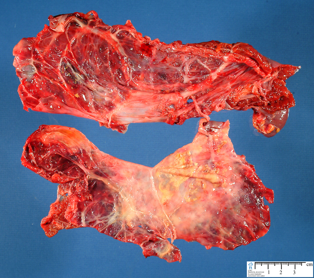
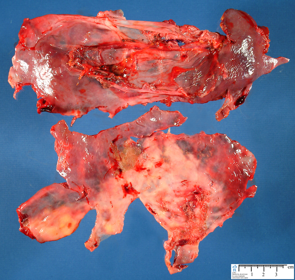
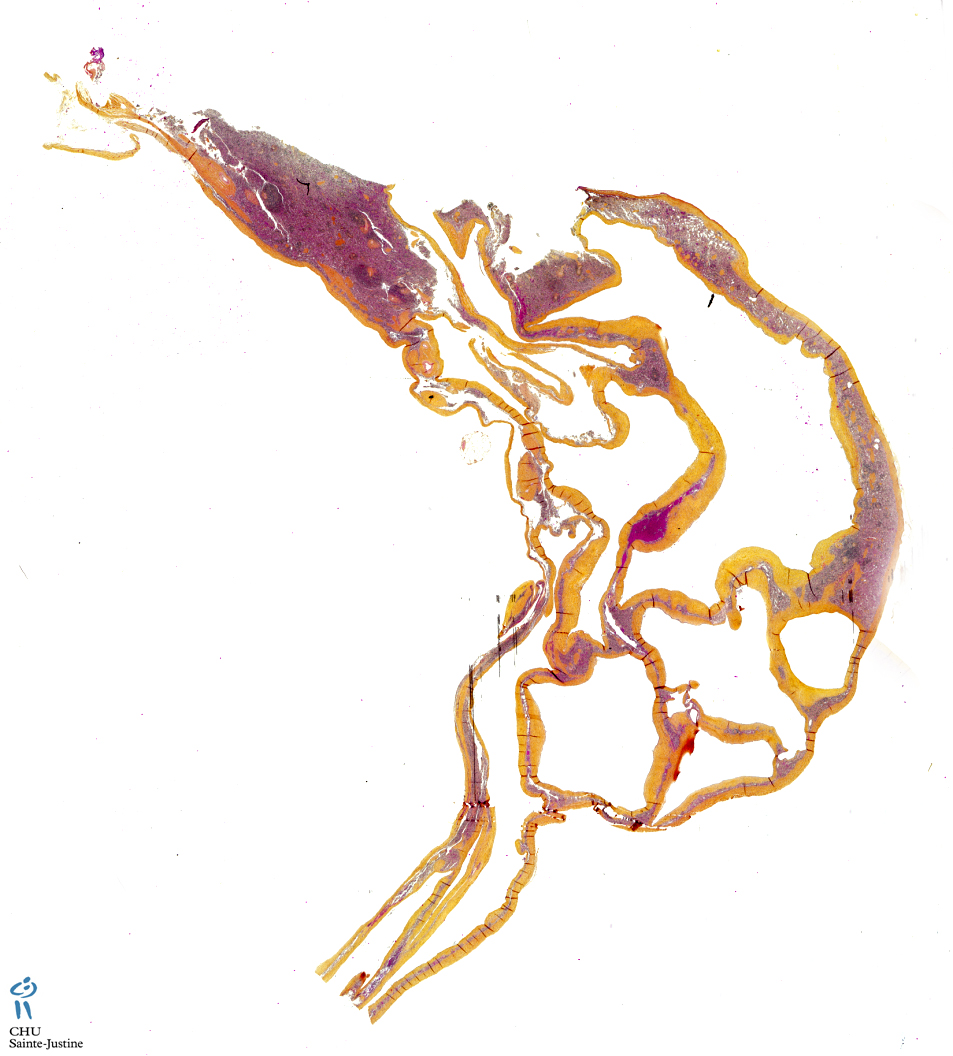
splenic mesothelial cyst
Image Gallery
[ (||image_reduire{0,60}|inserer_attribut{alt,Splenic mesothelial cyst}) ] [ (||image_reduire{0,60}|inserer_attribut{alt,Splenic mesothelial cyst (open)}) ] [ (||image_reduire{0,60}|inserer_attribut{alt,Splenic mesothelial cyst (open)}) ] [ (||image_reduire{0,60}|inserer_attribut{alt,Splenic multilocular mesothelial cysts}) ] [ (||image_reduire{0,60}|inserer_attribut{alt,Splenic multilocular mesothelial cysts}) ]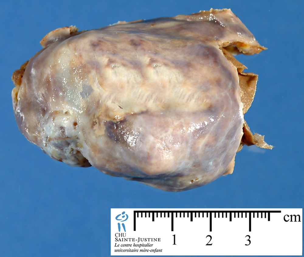
Synopsis
![]() cystic lesion
cystic lesion
- unifocal
- multifocal
![]() Size: microscopic size to 2 cm in diameter
Size: microscopic size to 2 cm in diameter
![]() thin-walled, semitranslucent, filled with clear or yellowish fluid or gelatinous contents
thin-walled, semitranslucent, filled with clear or yellowish fluid or gelatinous contents
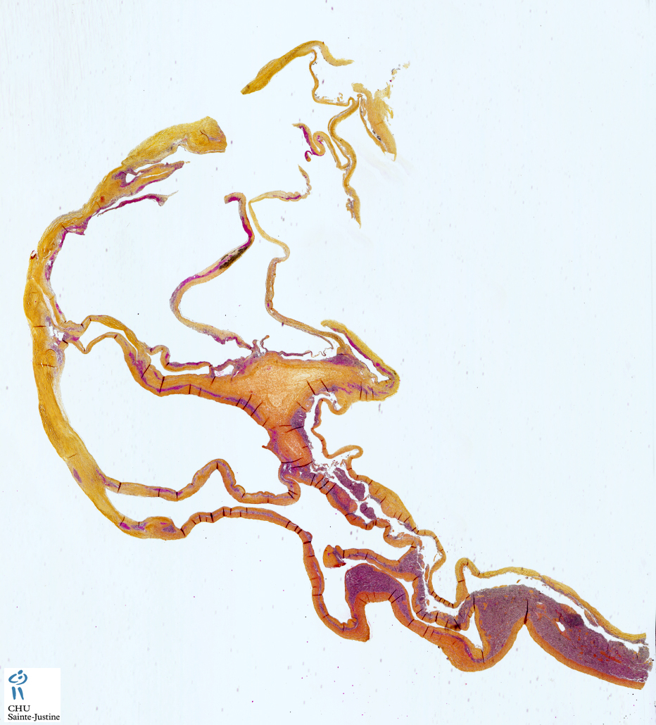
![]() cysts were with a single layer of flattened or cuboid mesothelial cells (CK+, calretinin+)
cysts were with a single layer of flattened or cuboid mesothelial cells (CK+, calretinin+)
![]() diffuse squamous cell metaplasia
diffuse squamous cell metaplasia
![]() cells focally forming small papillae
cells focally forming small papillae
![]() vacuolated cells
vacuolated cells
![]() no mucus
no mucus
Immunohistochemistry
![]() CK +
CK +
![]() calretin +
calretin +
Differential diagnosis
![]() splenic lymphangiomas (#9060604#)
splenic lymphangiomas (#9060604#)
![]() splenic lymphangiomatosis (#9060604#)
splenic lymphangiomatosis (#9060604#)
See also
![]() mesothelial cysts
mesothelial cysts
![]() diaphragmatic mesothelial cyst
diaphragmatic mesothelial cyst
![]() hepatic mesothelial cyst
hepatic mesothelial cyst
![]() splenic mesothelial cyst
splenic mesothelial cyst
References
![]() Arber DA, Strickler JG, Weiss LM. Splenic mesothelial cysts mimicking lymphagiomas. Am J Surg Pathol. 1997 Mar;21(3):334-8. PMID: #9060604#
Arber DA, Strickler JG, Weiss LM. Splenic mesothelial cysts mimicking lymphagiomas. Am J Surg Pathol. 1997 Mar;21(3):334-8. PMID: #9060604#