Home > D. General pathology > Blood and immunity > Spleen > spleen
spleen
Thursday 28 August 2003
Normal spleen. Adj. splenic
Digital slides
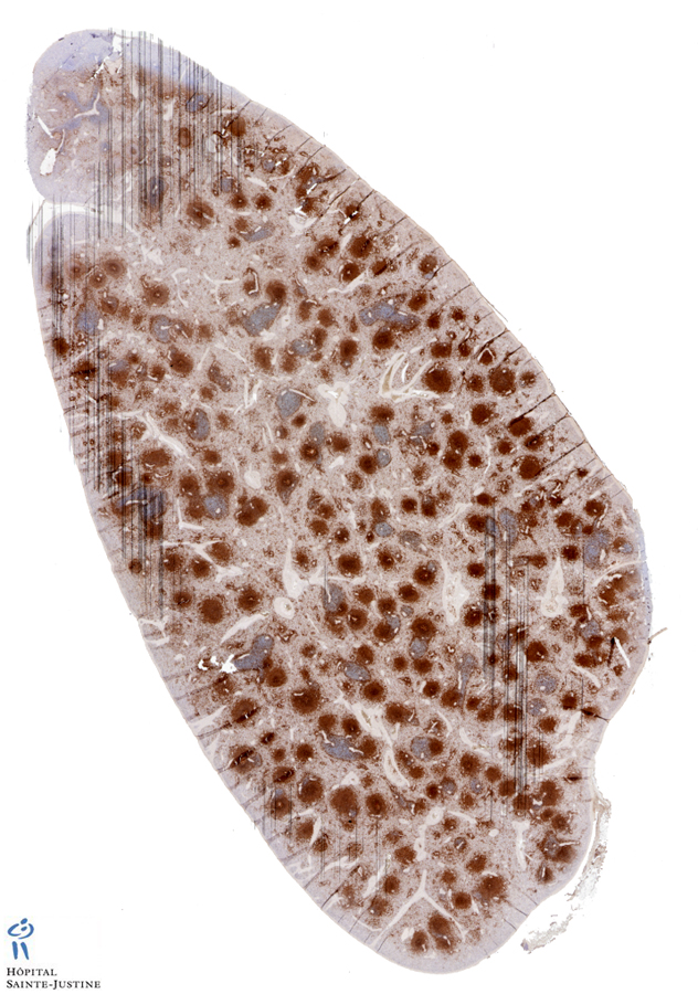
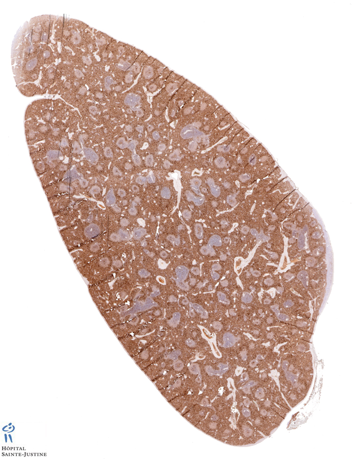
HPC:97 : Normal spleen
HPC:266 : Normal spleen
HPC:300 : Normal spleen (Idiopathic thrombopenic purpura)
Images
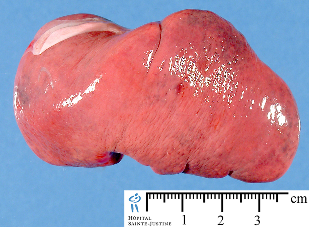
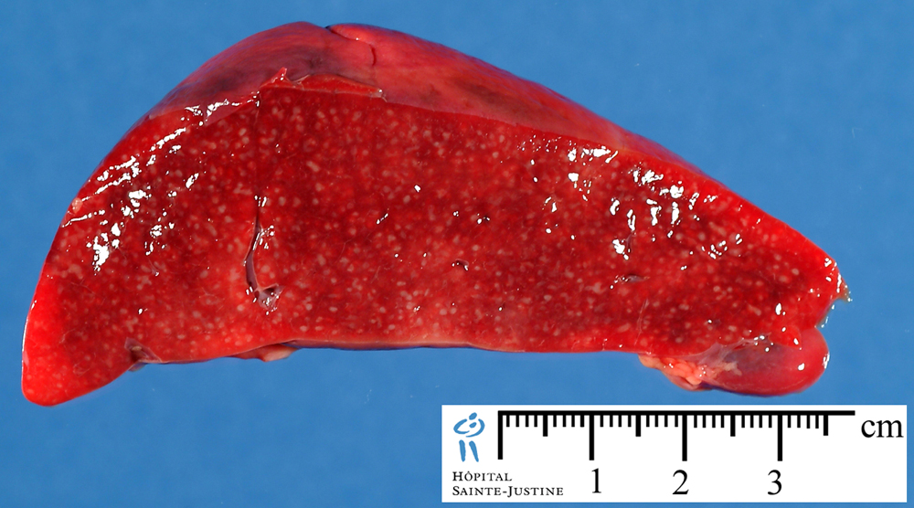
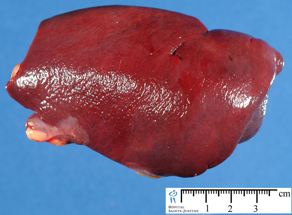
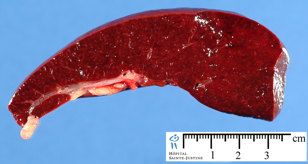
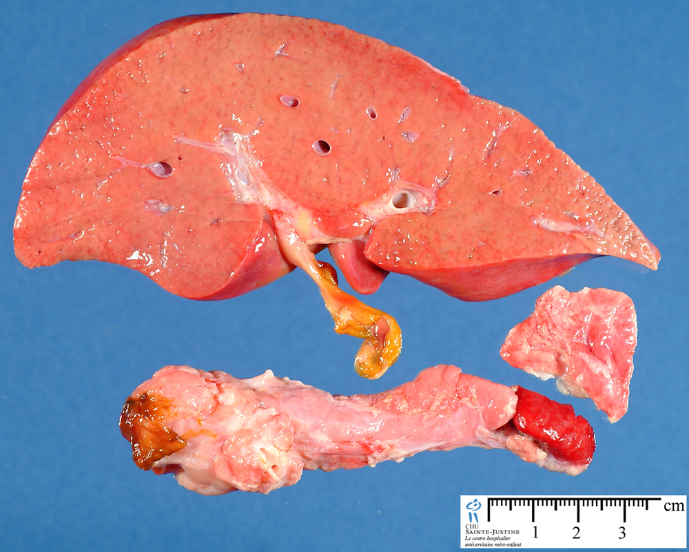
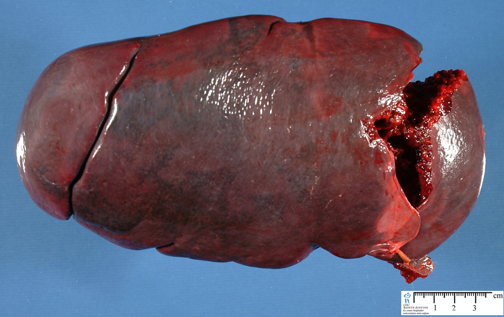
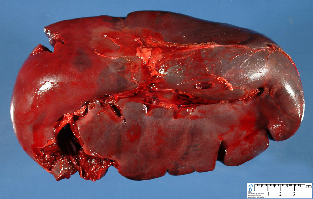
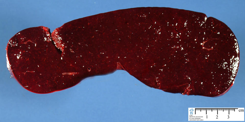
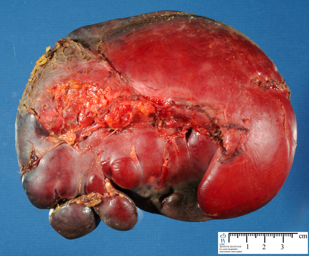
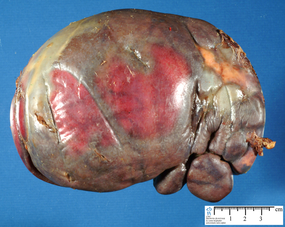
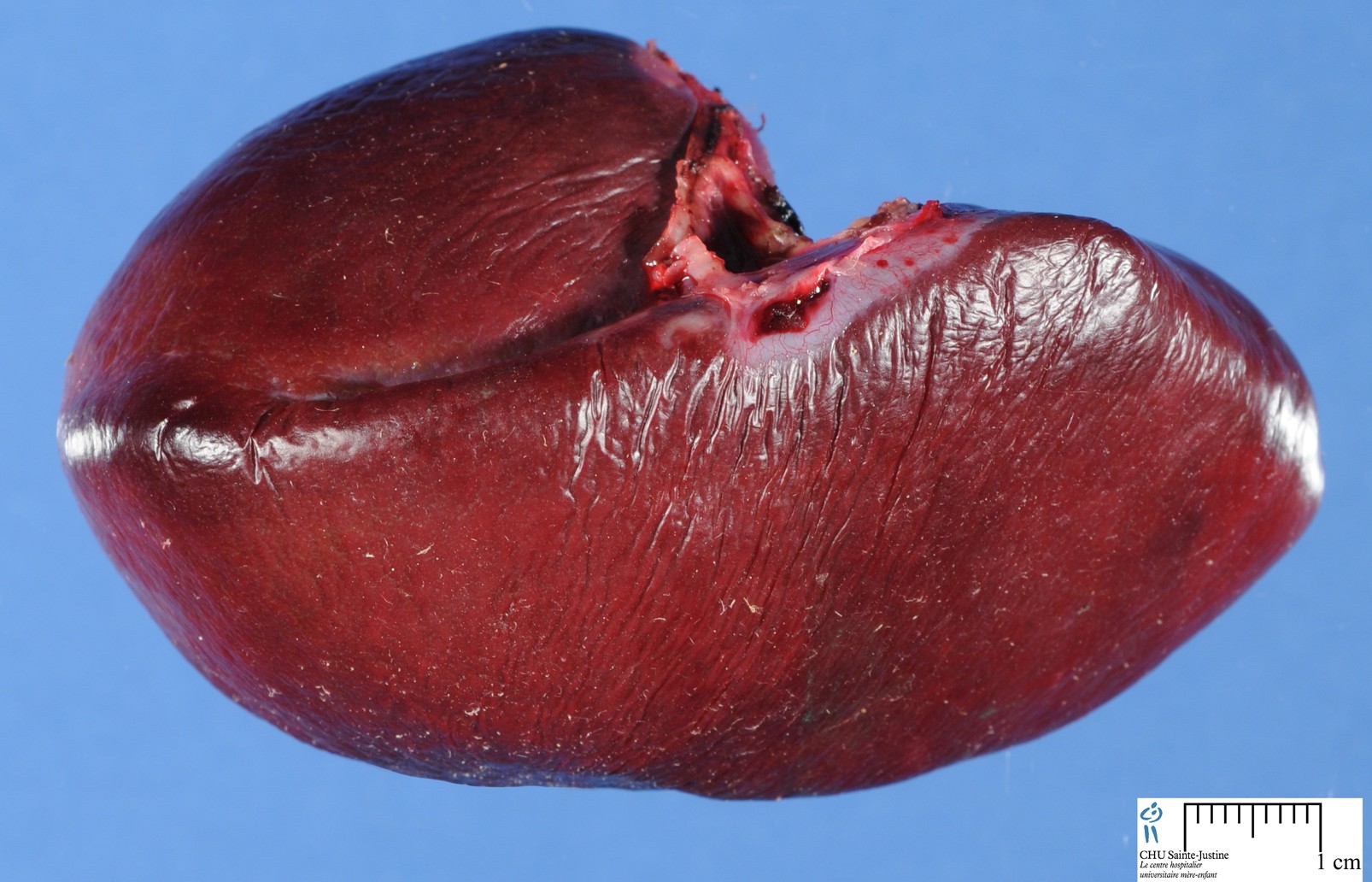
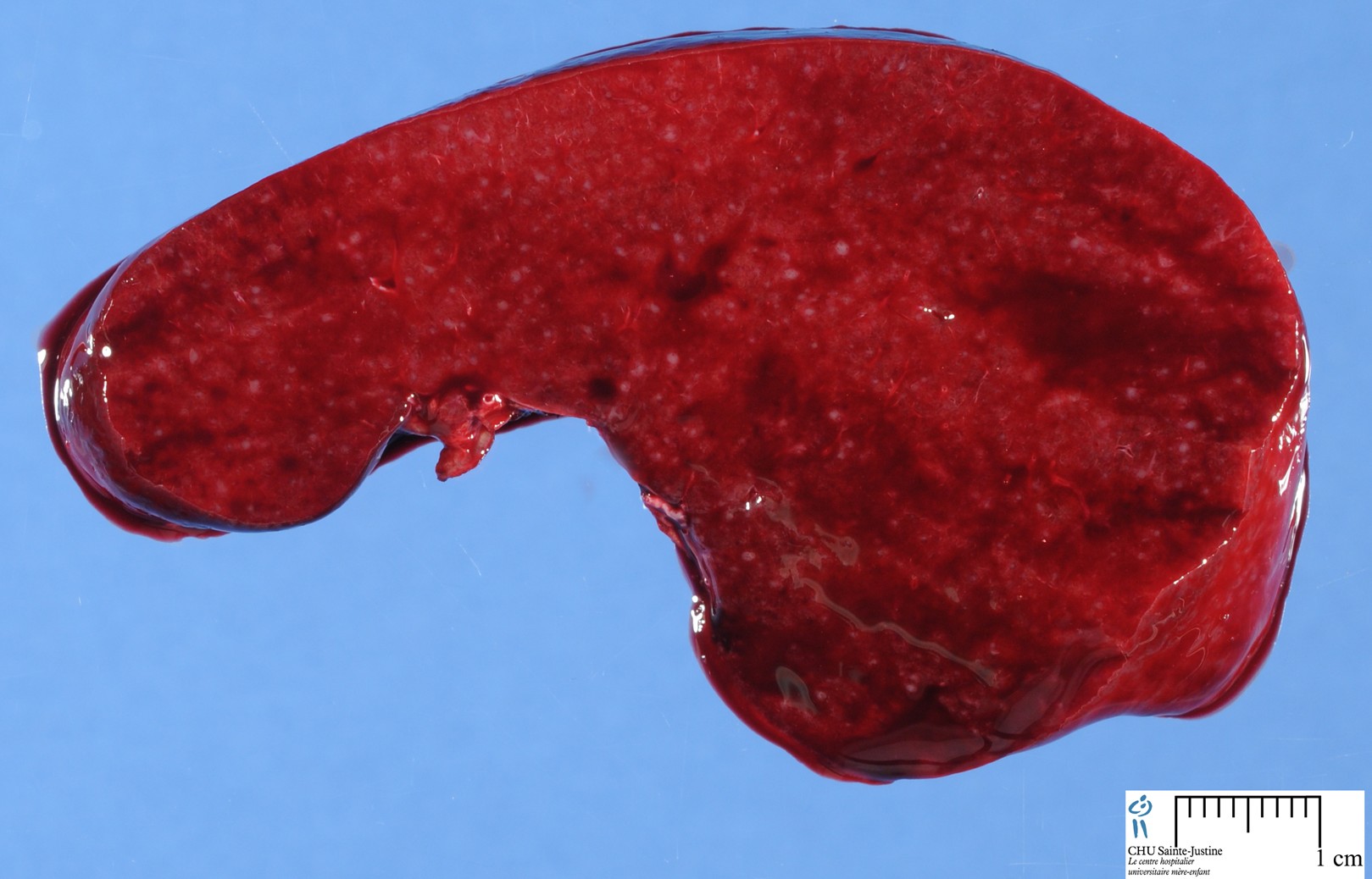
normal spleen
The spleen (from Greek σπλήν—splḗn) is an organ found in virtually all vertebrate animals. Similar in structure to a large lymph node, it acts primarily as a blood filter. Thus, life is possible after the spleen is removed.
The spleen is the largest secondary immune organ in the body and is responsible for initiating immune reactions to blood-borne antigens and for filtering the blood of foreign material and old or damaged red blood cells.
These functions are carried out by the 2 main compartments of the spleen, the white pulp (including the marginal zone) and the red pulp, which are vastly different in their architecture, vascular organization, and cellular composition.
Functions
The spleen plays important roles in regard to red blood cells (also referred to as erythrocytes) and the immune system. It removes old red blood cells and holds a reserve of blood in case of hemorrhagic shock and also recycles iron.
As a part of the mononuclear phagocyte system, it metabolizes hemoglobin removed from senescent erythrocytes. The globin portion of hemoglobin is degraded to its constitutive amino acids, and the heme portion is metabolized to bilirubin, which is removed in the liver.
The spleen synthesizes antibodies in its white pulp and removes antibody-coated bacteria and antibody-coated blood cells by way of blood and lymph node circulation.
A study published in 2009 using mice found that the spleen contains in its reserve half of the body’s monocytes within the red pulp. These monocytes, upon moving to injured tissue (such as the heart), turn into dendritic cells and macrophages while promoting tissue healing.
The spleen is a center of activity of the reticuloendothelial system and can be considered analogous to a large lymph node, as its absence causes a predisposition to certain infections.
Production of opsonins, properdin, and tuftsin.
Creation of red blood cells. While the bone marrow is the primary site of hematopoiesis in the adult, the spleen has important hematopoietic functions up until the fifth month of gestation. After birth, erythropoietic functions cease, except in some hematologic disorders.
As a major lymphoid organ and a central player in the reticuloendothelial system, the spleen retains the ability to produce lymphocytes and, as such, remains an hematopoietic organ.
Storage of red blood cells, lymphocytes and other formed elements. In horses, roughly 30% of the red blood cells are stored there. The red blood cells can be released when needed.
In humans, up to a cup (236.5 ml) of red blood cells can be held in the spleen and released in cases of hypovolemia. It can store platelets in case of an emergency. Up to a quarter of lymphocytes can be stored in the spleen at any one time.
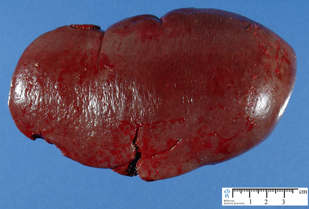
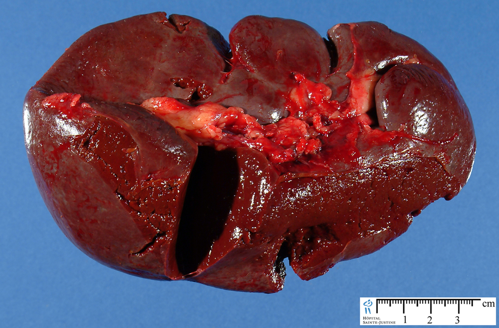
Macroscopy
In humans, the spleen is brownish in color and is located in the left upper quadrant of the abdomen. The spleen, in healthy adult humans, is approximately 11 centimetres (4.3 in) in length. It usually weighs between 150 grams (5.3 oz)[9] and 200 grams (7.1 oz).
The spleen is a dark red to blue-black organ located in the left cranial abdomen. It is adjacent to the greater curvature of the stomach and within the omentum. It is an elongated organ, roughly triangular in cross section. The gross appearance and size of the spleen are variable, depending on the species and the degree of distension; nonetheless, spleen weights can be important in its evaluation. The ratio of splenic weight to body weight remains fairly constant regardless of age and, in rats, is typically around 0.2%.
The spleen is surrounded by a capsule composed of dense fibrous tissue, elastic fibers, and smooth muscle. The outermost layer of the splenic capsule is composed of mesothelial cells, which may not be evident on histologic section. Irregularly spaced trabeculae of smooth muscle and fibroelastic tissue emanate from the capsule into the splenic parenchyma. These trabeculae also contain blood and lymph vessels and nerves. The lymph vessels are efferent vessels through which lymphocytes migrate to the splenic lymph nodes.
Histology - Components
splenic red pulp
- Red pulp is vascular, and has parencyhma and lots of vascular sinuses. These are sinuosoids - a specialised type of capillary, which is very leaky.
- The lining endothelial cells have wide slits between their lateral margins, that act as a filter. The blood cells have to move through these slits, before they can leave the spleen and worn out, or defective blood cells are damaged during this process. The damaged cells are then phagocytosed by the numerous macrophages in the red pulp, that lie just next to the sinusoids.
splenic white pulp
- White pulp contains lymphoid aggregations, mostly lymphocytes, and macrophages which are arranged around the arteries.
- The lymphocytes are both T (mainly T-helper) and B-cells.
splenic capsule and trabeculae
- The spleen is covered by a dense capsule, and there are connective tissue trabeculae, which provide internal support for the spleen, and carry the blood vessels into the spleen.
Pathology
splenic malformations
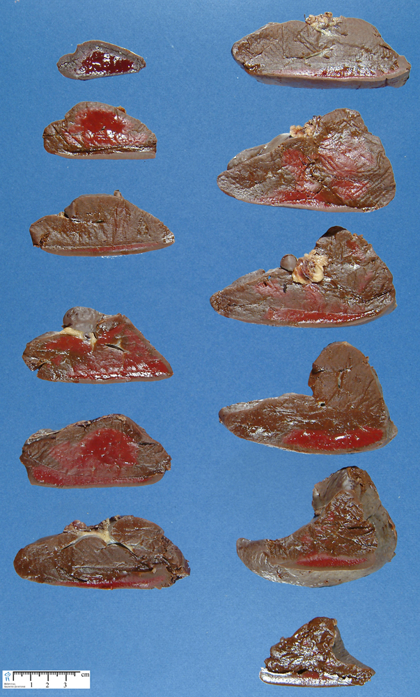
splenic anomalies (splenic lesions)
splenic diseases
splenic tumors
Videos
Histology of the spleen (by Washington deceit)
@<@object width="425" height="350">@<@param name="movie" value="http://www.youtube.com/v/Kl5L01BCYC8">@<@/param>@<@param name="wmode" value="transparent">@<@/param>@<@embed src="http://www.youtube.com/v/Kl5L01BCYC8" type="application/x-shockwave-flash" wmode="transparent" width="425" height="350">@<@/embed>@<@/object>
See also
Systems
- Organs