Onchocerca volvulus
Image Gallery
[ (||image_reduire{0,60}|inserer_attribut{alt,Microfilaria of Onchocerca volvulus, from skin snip.}) ]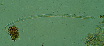
Digital slides
![]() UI:990 - Onchocerca volvulus
UI:990 - Onchocerca volvulus
Definition: Onchocerca volvulus is a filarial nematode transmitted by black flies. It affects more than 17 million people in Africa, South America, and Yemen.
An aggressive campaign of ivermectin treatment has dramatically reduced the incidence of Onchocerca infection in West Africa; however, O. volvulus remains the second most common preventable cause of blindness in sub-Saharan Africa.
Adult O. volvulus parasites mate in the dermis, where they are surrounded by a mixed infiltrate of host cells that produces a characteristic subcutaneous nodule (onchocercoma).
The major pathologic process, which includes blindness and chronic pruritic dermatitis, is caused by large numbers of microfilariae, released by females, that accumulate in the skin and in the eye chambers. Punctate keratitis is caused by inflammation around a degenerating microfilaria.
It is sometimes accentuated by treatment with antifilarial drugs (Mazzotti reaction), resulting in blindness. Ivermectin kills only immature worms, not adult worms, so parasites repopulate the host a few months after treatment. Recently, doxycycline treatment has been shown to block reproduction of O. volvus for up to 24 months.
Doxycycline kills Wolbachia, which are symbiotic bacteria that live inside adult O. volvulus and are required for worm fertility, similar to filarial nematodes.
O. volvulus causes chronic, itchy dermatitis with focal darkening or loss of pigment and scaling, referred to as leopard, lizard, or elephant skin. Foci of epidermal atrophy and elastic fiber breakdown may alternate with areas of hyperkeratosis, hyperpigmentation with pigment incontinence, dermal atrophy, and fibrosis.
The subcutaneous onchocercoma is composed of a fibrous capsule surrounding adult worms and a mixed chronic inflammatory infiltrate that includes fibrin, neutrophils, eosinophils, lymphocytes, and giant cells.
The progressive eye lesions begin with punctate keratitis along with small, fluffy opacities of the cornea caused by degenerating microfilariae, which evoke an eosinophilic infiltrate.
This is followed by a sclerosing keratitis that opacifies the cornea, beginning at the scleral limbus. Microfilariae in the anterior chamber cause iridocyclitis and glaucoma, whereas involvement of the choroid and retina results in atrophy and loss of vision.
See also
![]() onchocercosis
onchocercosis
![]() filariasis
filariasis
![]() nematodosis
nematodosis
![]() helminthiasis
helminthiasis