Home > E. Pathology by systems > Respiratory system > Lungs > pulmonary carcinomas
pulmonary carcinomas
Tuesday 25 May 2004
lung carcinomas, lung cancers, lung cancer
Digital case
JRC:1593 : Pulmonary adenocarcinoma arinsing in a sequestred lung (pulmonary sequestration).
JRC:1594 : Pulmonary small cell adenocarcinoma.
JRC:1602 : Pulmonary squamous cell carcinoma (variable differentiation).
JRC:1596 : Bronchioloalveolar carcinoma.
JRC:6222 : pulmonary giant cell carcinoma.
Epidemiology
Lung cancer is the most common cancer worldwide, with an estimated 1,600,000 new cases and 1,380,000 deaths in 2008.
Classification
Classification of lung carcinomas by histopathologic subtype provides important information about prognosis and is necessary for optimal treatment. Traditionally, the classification of lung carcinoma has been based solely on evaluation of routinely stained biopsies or cytologic preparations. Increasingly, however, ancillary tests such as immunohistochemistry are being used to aid pathologists in diagnosis of subtypes.
IASLC 2011 - In 2011, a multidisciplinary expert panel representing the International Association for the Study of Lung Cancer (IASLC), the American Thoracic Society (ATS), and the European Respiratory Society (ERS) proposed a major revision of the classification system. These changes primarily affect the classification of adenocarcinoma and its distinction from squamous cell carcinoma.
The rationale for these IASLC/ATS/ERS recommendations is based upon several observations:
Advances in understanding the specific molecular pathways that drive malignancy have opened up new pathways for treatment, and molecular characterization of patients with adenocarcinoma is resulting in the use of agents with high levels of antitumor activity. As an example, epidermal growth factor receptor (EGFR) tyrosine kinase inhibitors are the preferred initial treatment for patients with metastatic lung adenocarcinoma whose tumors have characteristic activating mutation in EGFR. Targeted therapy is also being developed for those with adenocarcinomas whose tumors harbor the EML4-ALK translocation. (See "Targeted agents in the initial systemic treatment of advanced non-small cell lung cancer", section on ’EGFR TK inhibitors’ and "EML4-ALK fusion oncogene positive advanced non-small cell lung cancer".)
Distinguishing squamous carcinoma from other non-small carcinomas, particularly adenocarcinoma, is important for patients with advanced stage disease as certain chemotherapeutic agents are contraindicated for patients with squamous histology. The 2004 WHO classification did not address how this distinction was to be made on small biopsies and specifically endorsed lumping tumors under the “non small cell carcinoma” rubric. The new classification scheme offers guidelines for how to use a panel of immunohistochemical stains (eg, p63, TTF-1, cytokeratin 5/6) when obvious features of a specific line of differentiation are not apparent on a small biopsy.
Radiographic abnormalities, particularly the appearance of small carcinomas with a ground glass appearance, help to define a subset of patients with adenocarcinoma, who have an excellent prognosis with complete surgical resection. Less extensive surgery may be adequate treatment in such cases.
The following discussion incorporates that proposed revision and provides correlation with the WHO schema.
Types
pulmonary adenocarcinoma
pulmonary squamous cell carcinoma
pulmonary pleomorphic carcinoma
pulmonary mucinous carcinoma
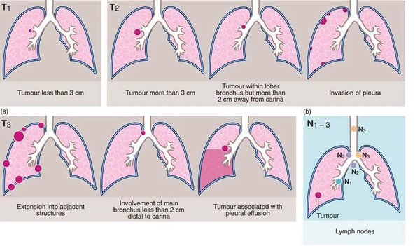
TNM staging : lung cancer TNM staging system 2009
New TNM classification system: http://staginglungcancer.org
T1: Tumor @<@ 3 cm diameter, surrounded by lung or visceral pleura, without invasion more proximal than lobar bronchus.
T1a @<@ 2 cm
T1b @<@ 3 cm
T2: Tumor > 3 cm but @<@ 7 cm, or tumor with any of the following features:
Involves main bronchus > 2 cm distal to carina
Invades visceral pleura
Associated with atelectasis or obstructive pneumonitis that extends to the hilar region but does not involve the entire lung
T3: Tumor > 7 cm or any of the following:
Directly invades any of the following: chest wall, diaphragm, phrenic nerve, mediastinal pleura, parietal pericardium, main bronchus @<@ 2 cm from carina without involvement of the carina.
Atelectasis or obstructive pneumonitis of the entire lung
Separate tumor nodules in the same lobe
T4: Tumor of any size that invades the mediastinum, heart, great vessels, trachea, recurrent laryngeal nerve, esophagus, vertebral body, carina, or with separate tumor nodules in a different ipsilateral lobe.
(Reference: http://www.atcs.jp/pdf/2009_15_1/4.pdf)
Familial susceptibility loci
6q23-25 (15272417)
Oncogenetics
51 genomic regions with homozygous deletions (HDs) (17674361)
Fusion genes
EML4/ALK fusion gene in non-small-cell lung cancer (17625570)
Reviews
Personalized medicine for lung cancer: new challenges for pathology. Kerr KM. Histopathology. 2012 Mar;60(4):531-46. PMID: 21916947
Molecular diagnostics of lung carcinomas. Dacic S. Arch Pathol Lab Med. 2011 May;135(5):622-9. PMID: 21526960
Lung cancer and the future of pathology. Cagle PT, Dacic S. Arch Pathol Lab Med. 2011 Mar;135(3):293-5. PMID: 21366449
Revolution in lung cancer: new challenges for the surgical pathologist. Cagle PT, Allen TC, Dacic S, Beasley MB, Borczuk AC, Chirieac LR, Laucirica R, Ro JY, Kerr KM. Arch Pathol Lab Med. 2011 Jan;135(1):110-6. PMID: 21204716
Targeted Therapies in Lung Cancer. Chirieac LR, Dacic S. Surg Pathol Clin. 2010 Mar 1;3(1):71-82. PMID: 20680095
Molecular testing in lung carcinoma: Quo vadis? Dacic S, Yousem SA. Am J Clin Pathol. 2010 Jul;134(1):7-9. PMID: 20551260
Sun S, Schiller JH, Gazdar AF. Lung cancer in never smokers—a different disease. Nat Rev Cancer. 2007 Oct;7(10):778-90. PMID: 17882278
Wistuba II. Genetics of preneoplasia: lessons from lung cancer. Curr Mol Med. 2007 Feb;7(1):3-14. PMID: 17311529
Meuwissen R, Berns A. Mouse models for human lung cancer. Genes Dev. 2005 Mar 15;19(6):643-64. PMID: 15769940
References
Soda M, Choi YL, Enomoto M, Takada S, Yamashita Y, Ishikawa S, Fujiwara SI, Watanabe H, Kurashina K, Hatanaka H, Bando M, Ohno S, Ishikawa Y, Aburatani H, Niki T, Sohara Y, Sugiyama Y, Mano H. Identification of the transforming EML4-ALK fusion gene in non-small-cell lung cancer. Nature. 2007 Jul 11; PMID: 17625570
Nagayama K, Kohno T, Sato M, Arai Y, Minna JD, Yokota J. Homozygous deletion scanning of the lung cancer genome at a 100-kb resolution. Genes Chromosomes Cancer. 2007 Aug 2; PMID: 17674361