Home > E. Pathology by systems > Genital system > Female genital system > Breast > collagenous spherulosis
collagenous spherulosis
Thursday 16 June 2016
Mucinous spherulosis
Spherulosis
Adenoid cystic hyperplasia
Definition: Collagenous spherulosis” (CS) is an uncommon benign change (occurs in less than 1-2% of all biopsies) and is an incidental microscopic finding involving lobular acini and ductules. It consists of intra-luminal clusters of eosinophilic spherules.
CS is considered as an end-stage lesion resulting from transformation of its predecessor, mucinous spherulosis of the early stages.
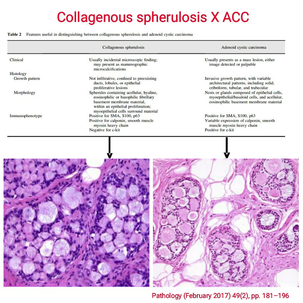
Collagenous spherulosis usually does not form a breast mass, it is a microscopic finding. It is positive for smooth muscle markers and negative for CD117, and has PAS positive basement membrane material.
Images
Collagenous spherulosis
- https://www.ncbi.nlm.nih.gov/pmc/articles/PMC3927328/figure/F1/
- https://twitter.com/FKhaniPath/status/836423832000937984
- https://twitter.com/TheUSCAP/status/706658202616799232
- https://twitter.com/TheUSCAP/status/706658202616799232
LCIS arising within collagenous spherulosis
atypical lobular hyperplasia - ALH involving collagenous spherulosis
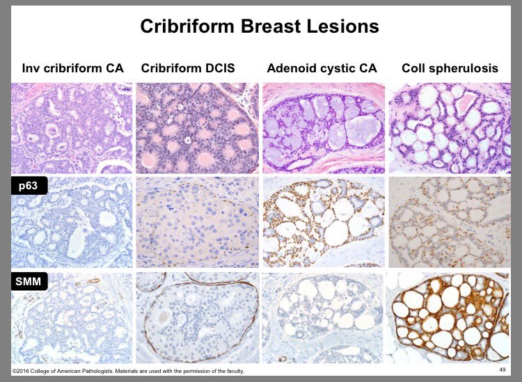
Collagenous spherulosis vs adenoid cystic carcinoma
LCIS involving collagenous spherulosis
- https://twitter.com/LizaMQuintana/status/844284323297660932
- https://twitter.com/angeldeoromx/status/755567054808166400
Sclerosing papilloma (mimics invasive carcinoma) with collagenous spherulosis (mimics DCIS).
Microscopy
On histologic examination, CS is characterized by presence of eosinophilic intra-luminal collagen rich spherules measuring 20-100 microns in diameter, surrounded by flattened myoepithelial cells.
Most frequently, it shows a central floccular aggregate with radiating spikes that merge with the scalloped projections at the periphery. The material may also be finely granular and more evenly distributed. The periphery of the sphere may be marked by an eosinophilic cuticle of variable thickness and staining intensity. Sometimes the cuticle is associated with a myoepithelial cell nucleus.
As many as 50 spherules can be seen per section of the lesion and are usually discrete but may coalesce. It was shown that the hyaline material present within the intraluminal space was rich in collagen by conventional histochemistry, and was subsequently proved by immunocytochemistry that it was one of the component of the basement membrane.
Associations
CS has been described in association with various benign and malignant lesions, including sclerosing adenosis , radial scar , intraductal papilloma , fibroadenoma , atypical ductal hyperplasia , ductal carcinoma in situ ( DCIS ) and lobular carcinoma in situ ( LCIS ) of the breast.
Salivary glands
CS has been seen in salivary gland tumors like sclerosing polycystic adenosis , epithelial-myoepithelial carcinoma , polymorphous low grade adenocarcinoma and cutaneous myoepithelial tumors .
Physiopathology
The most accepted theory on the mechanism of spherule formation is the secretion of extracellular material by the proliferative myoepithelium.
Based on the observations of a circumscribed spherule with frequent identification of a compressed myoepithelial cell nucleus surrounding collagenous spherulosis, the spherules can be interpreted to be formed by extracellular material deposition.
The finding of laminin and type IV collagen in the spherules within “collagenous spherulosis” suggests that the spherules contain basement membrane material. In some instances, the contents of the spherule can undergo calcification (25% of cases of collagenous spherulosis have associated calcifications)
See also
- mammary epithelial anomalies
Open references
Collagenous spherulosis. Bavle RM. J Oral Maxillofac Pathol. 2013 Sep;17(3):322-3. doi : 10.4103/0973-029X.125174 PMID: 24574645 Free
Paywall references
Lobular Carcinoma In Situ with Collagenous Spherulosis: Clinicopathologic Characteristics of 38 Cases. 2017. doi : 10.1111/tbj.12292