Home > G. Tumoral pathology > juvenile xanthogranuloma
juvenile xanthogranuloma
Friday 24 October 2003
juvenile xanthogranulomas; juvenile xanthogranuloma - generalized eruptive histiocytosis
Definition: Juvenile xanthogranuloma (JXG), or generalized eruptive histiocytosis, is a rare disorder of the non-Langerhans cell histiocytosis (LCH) type with unknown etiology and pathogenesis. It is still unclear whether JXG is a reactive or a neoplastic process. Clonality has been assessed (17460468). It can be localized or multicentric.
Images
https://twitter.com/GSomersPath/status/1163452727567015936
https://twitter.com/histiocytosisX/status/886280528973922304
https://twitter.com/LizMontgomeryMD/status/1142192420731273217
https://twitter.com/path_tips/status/1009806871631364098
https://twitter.com/DermpathUAMS/status/500461196781563904
https://twitter.com/slusagar/status/1481642200572256256
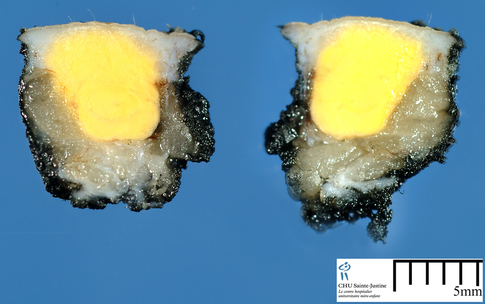
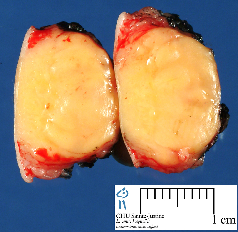
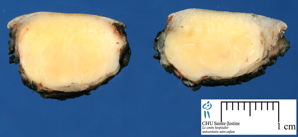
Juvenile xanthogranuloma is frequently found on the head and trunk in infants, but can be diagnosed in older patients. It is composed of a dense lymphohistiocytic proliferation that stains positive for CD68 and vimentin.
XG inflammation is a specific and unusual histopathologic entity that can affect several organs. It has been encountered predominantly in the kidney and gallbladder, and less frequently in the urinary bladder, endometrium, fallopian tube, ovary, vagina, prostate, testis, epididymis, colon, and appendix.
Involvement of the brain, lungs, or bone is very rare.
The age at presentation in these reports ranged from the 2nd to the 6th decade with a male predominance.
The exact etiology is unclear, but delayed-type hypersensitivity may be implicated in the pathogenesis. Complete surgical excision appears to be curative.
No recurrences have been reported.
JXG is a proliferative disorder of dendrocytes, possibly dermal dendrocytes. Juvenile xanthogranulomas is a histiocytic disorder, primarily but not exclusively seen throughout the first two decades of life and principally as a solitary cutaneous lesion.
Juvenile xanthogranuloma (JXG) and LCH have been grouped in the category of dendritic cell-related disorders and separated from macrophage-related proliferations like the Rosai-Dorfman disease.
Despite the supposed similarities between JXG and LCH in terms of their histogenesis, they differ in their morphology and immunophenotype, as well as in their clinical presentation. These differences might explain the poorer outcome of LCH compared with the excellent prognosis of JXG.
JXG is a clonal proliferation of histiocytic/dendritic cells and does not differ in this respect from LCH.
Images
Juvenile xanthogranuloma and Touton cells
- https://twitter.com/histiocytosisX/status/886280528973922304
- https://twitter.com/DanglisFotiosMD/status/867983078236463104
- https://twitter.com/NejibY/status/893195720244813826
- https://twitter.com/PathologyTweet/status/870628660964995073
- https://twitter.com/Dermpathl/status/830899773330821120
- https://twitter.com/RoshanNasim/status/813811749891620868
- https://twitter.com/MegKetchamMD/status/791635082452885504
- https://twitter.com/BeynonMD/status/735623896737583105
Juvenile xanthogranuloma (CD68+/S100-/CD1a-) with unusual nuclear factor XIIIA positivity (clone AC-1A1).
Digital cases
JRC:11021 : Juvenile xanthogranuloma.
Presentation (12717244)
solitary cutaneous lesion (67%)
solitary subcutaneous or deep soft tissue mass (16%)
multiple cutaneous lesions (7%)
solitary extracutaneous, nonsoft tissue lesion (5%)
multiple cutaneous and visceral-systemic lesions (5%)
systemic juvenile xanthogranuloma (17230073)
macrophagic activation syndrome (MAS)
Localization
cutis and subcutis (cutaneous juvenile xanthogranuloma)
mouth, oral cavity (12324792)
peripheral nerve (11257628)
heart (11178674)
eyes (6819503): choroid (12107521), iris (9514494, 17317408), orbit (2123087)
central nervous system (3082946), brain (9485160), intracranial (17332947)
liver (12717244)
spleen
lung
oropharynx
muscle
bone : osseous juvenile xanthogranuloma / JXG of the bone (17378689)
Variants
solitary juvenile xanthogranuloma
multiple juvenile xanthogranuloma
systemic juvenile xanthogranuloma (8765620)
nonlipidized juvenile xanthogranuloma (9144693)
mitotically active juvenile xanthogranuloma (12296763)
Juvenile xanthogranuloma group histiocytic disorders (dermal dendrocyte)
progressive nodular histiocytosis
xanthoma disseminatum
benign cephalic histiocytosis
spindle cell xanthogranuloma
generalized eruptive histiocytosis
Microscopical synopsis
Touton giant cell (consistent in cutaneous lesions; absent or present in reduced numbers in extracutaneous lesions)
spindle cells intermingled among the mononuclear cells or forming short fascicles
Immunohistochemistry
vimentin+
CD68+
factor XIIIa+
fascin+
alpha-1-antitrypsine
S-100-
CD1a-
Predisposition
neurofibromatoses
- neurofibromatosis type 1 (NF1) (15078345, 15342987)
- neurofibromatosis type 2 (NF2) (9810909)
Associations
neurilemmomatosis (multiple neurilemmomas) (9810909)
cafe-au-lait spots (CALS) (11454088, 15078345)
prolonged severe pancytopenia (16047346)
juvenile myelomonocytic leukemia (juvenile myeloid leukemia, juvenile chronic myelogenous leukaemia) (6422862, 14550978) or acute myelomonocytic leukemia (6430255) -* with neurofibromatosis type 1 (NF1) (15342987, 14550978)
Clonality
Monoclonality has been assessed in one case (17460468).
Cytogenetics: no data
Molecular biology: no data
Differential diagnosis
histiocytic proliferations (histiocytoses)
- dendritic cell-related proliferations
- Langerhans cell histiocytosis
- polyclonal proliferations of macrophages
- Rosai-Dorfmann disease
See also
xanthogranulomas
histiocytic infiltration
histiocytoses
histiocytes
References
Janssen D, Harms D. Juvenile xanthogranuloma in childhood and adolescence: a clinicopathologic study of 129 patients from the kiel pediatric tumor registry. Am J Surg Pathol. 2005 Jan;29(1):21-8. PMID: 15613853
Dehner LP. Juvenile xanthogranulomas in the first two decades of life: a clinicopathologic study of 174 cases with cutaneous and extracutaneous manifestations. Am J Surg Pathol. 2003 May;27(5):579-93. PMID: 12717244
Hu WK, Gilliam AC, Wiersma SR, Dahms BB. Fatal congenital systemic juvenile xanthogranuloma with liver failure. Pediatr Dev Pathol. 2004 Jan-Feb;7(1):71-6. PMID: 15255037