Synopsis
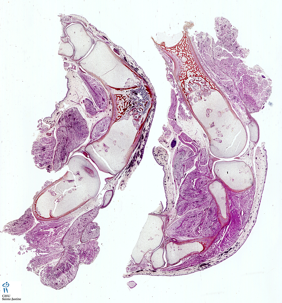
![]() extreme disorganization and retardation of the physeal growth zone
extreme disorganization and retardation of the physeal growth zone
![]() in achondrogenesis type 1A (IA), resting chondrocytes frequently contain characteristic large PAS-positive, diastase resistant cytoplasmic inclusions (spherical or oval, lying within membrane-bound vacuoles) (chondrocytic inclusions)
in achondrogenesis type 1A (IA), resting chondrocytes frequently contain characteristic large PAS-positive, diastase resistant cytoplasmic inclusions (spherical or oval, lying within membrane-bound vacuoles) (chondrocytic inclusions)
![]() in achondrogenesis type 1B (IB), resting cartilage is characterized by matrix deficiency and perichondrocytic collagen rings which stain trichrome, silver methenamine and toluidine blue. Chondrocytic inclusions are absent.
in achondrogenesis type 1B (IB), resting cartilage is characterized by matrix deficiency and perichondrocytic collagen rings which stain trichrome, silver methenamine and toluidine blue. Chondrocytic inclusions are absent.
- generalized deficiency of the matrix
- increased number of markedly enlarged lacunae and chondrocytes that may have abundant clear cytoplasm
- cartilage canals are markedly enlarged, stellate in shape and fibrotic
- the physeal growth zone is markedly retarded and disorganized with closely arranged large chondrocytic lacunae and prominently deficient intervening matrix
- predominant collagen is type 1 rather than type 2 (disorder of type 2 collagen biosynthesis by COL2A1 germline mutations)
Types
![]() achondrogenesis type 1 (AGC1s)
achondrogenesis type 1 (AGC1s)
- achondrogenesis type 1A (AGC1A) (MIM.200600)
- achondrogenesis type 1B (AGC1B) (SLC26A2 at 5q32-q33.1) (MIM.600972)
![]() achondrogenesis type 2 (AGC2) (COL2A1 at 12q13.11-q13.2) (MIM.200610)
achondrogenesis type 2 (AGC2) (COL2A1 at 12q13.11-q13.2) (MIM.200610)
![]() achondrogenesis type 3 (AGC3) (MIM.200710)
achondrogenesis type 3 (AGC3) (MIM.200710)
![]() achondrogenesis type 4 (AGC4) (MIM.200720)
achondrogenesis type 4 (AGC4) (MIM.200720)
Case records
![]() Case 10610: Achondrogenesis type 2
Case 10610: Achondrogenesis type 2
See also