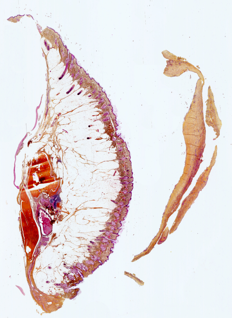
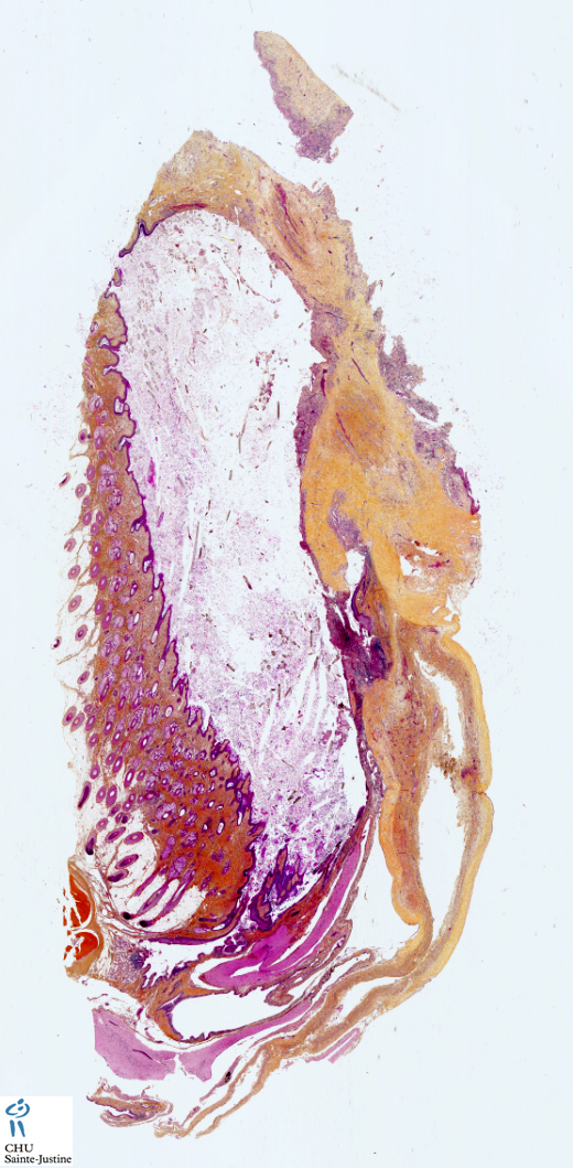
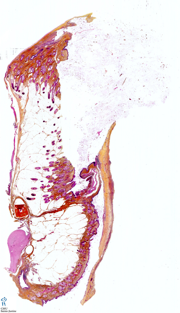
ovarian mature cystic teratoma
Image Gallery
[ (||image_reduire{0,60}|inserer_attribut{alt,Ovarian mature cystic teratoma}) ] [ (||image_reduire{0,60}|inserer_attribut{alt,Ovarian mature cystic teratoma}) ] [ (||image_reduire{0,60}|inserer_attribut{alt,Ovarian mature cystic teratoma}) ] [ (||image_reduire{0,60}|inserer_attribut{alt,Ovarian mature cystic teratoma}) ]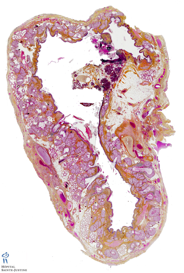
Definition: Mature cystic teratomas (MCTs) of the ovary are typically composed of a cyst lined with neoplastic tumor cells recapitulating ectodermal differentiation including derivatives such as epidermis, dermis, and appendages (sebaceous glands), although mesodermal and endodermal derivatives can also be seen.
Variants
![]() oligodendroglioma arising in OMCT (#19483626#)
oligodendroglioma arising in OMCT (#19483626#)
![]() chordoma arising in OMCT (#17418959#)
chordoma arising in OMCT (#17418959#)
Components
![]() Meningothelial tissue
Meningothelial tissue
- Meningothelial proliferation similar to ectopic meningothelial hamartoma (EMH) can also been observed.
- The presence of meningothelial tissue in MCT of the ovary is quite frequent and its appearance is similar to that of ectopic meningothelial hamartoma (EMH).
- The similar morphologic appearance of the meningothelial proliferation in MCT to EMH, its localization to the dermal subcutaneous portion of MCT, and its frequent proximity to glial tissue supports the hypothesis that the tissue elements of ovarian MCT are primarily of the head and neck type (eg, scalp skin, brain, upper respiratory/sinonasal, and less commonly thyroid).
- It suggests that the neoplastic growth of MCT parallels normal anterior embryonic plate development with primarily the formation of the cranial (cephalad) portion of the embryo.
Cytogenetics
Mature ovarian teratomas are benign ovarian germ cell tumors that usually present with a normal karyotype.
Rarely:
![]() monosomies of chromosomes 6, 14, 16, and 21
monosomies of chromosomes 6, 14, 16, and 21
![]() trisomies of chromosomes 14 and 21
trisomies of chromosomes 14 and 21
![]() deletions of Xq, 5p, 16p, and 17p.
deletions of Xq, 5p, 16p, and 17p.
Case records
See also
![]() teratomas
teratomas
- ovarian teratoma
- ovarian dermoid cyst
References
![]() Meningothelial proliferations in mature cystic teratoma of the ovary: evidence for the common presence of cranially derived tissues paralleling anterior embryonic plate development. An analysis of 25 consecutive cases. Chen E, Fletcher CD, Nucci MR. Am J Surg Pathol. 2010 Jul;34(7):1014-8. PMID: #20534997#
Meningothelial proliferations in mature cystic teratoma of the ovary: evidence for the common presence of cranially derived tissues paralleling anterior embryonic plate development. An analysis of 25 consecutive cases. Chen E, Fletcher CD, Nucci MR. Am J Surg Pathol. 2010 Jul;34(7):1014-8. PMID: #20534997#
![]() Oligodendroglioma arising in an ovarian mature cystic teratoma. Opris I, Ducrotoy V, Bossut J, Lamy A, Sabourin JC. Int J Gynecol Pathol. 2009 Jul;28(4):367-71. PMID: #19483626#
Oligodendroglioma arising in an ovarian mature cystic teratoma. Opris I, Ducrotoy V, Bossut J, Lamy A, Sabourin JC. Int J Gynecol Pathol. 2009 Jul;28(4):367-71. PMID: #19483626#
![]() Chordoma arising in a mature cystic teratoma of the ovary: a case report. Las Heras F, Pritzker KP, Colgan TJ. Pathol Res Pract. 2007;203(6):467-71. PMID: #17418959#
Chordoma arising in a mature cystic teratoma of the ovary: a case report. Las Heras F, Pritzker KP, Colgan TJ. Pathol Res Pract. 2007;203(6):467-71. PMID: #17418959#
![]() Comprehensive cytogenetic evaluation of a mature ovarian teratoma case. Schmid-Braz AT, Cavalli LR, Cornélio DA, Wuicik L, Ribeiro EM, Bleggi-Torres LF, Lima RS, de Andrade Urban C, Haddad BR, Cavalli IJ. Cancer Genet Cytogenet. 2002 Jan 15;132(2):165-8. PMID: #11850083#
Comprehensive cytogenetic evaluation of a mature ovarian teratoma case. Schmid-Braz AT, Cavalli LR, Cornélio DA, Wuicik L, Ribeiro EM, Bleggi-Torres LF, Lima RS, de Andrade Urban C, Haddad BR, Cavalli IJ. Cancer Genet Cytogenet. 2002 Jan 15;132(2):165-8. PMID: #11850083#