mixed germ cell tumor
Image Gallery
[ (||image_reduire{0,60}|inserer_attribut{alt,Chondroid teratoma component of a mixed germ cell tumor (yolk sac (...)}) ] [ (||image_reduire{0,60}|inserer_attribut{alt,Chondroid teratoma component of a mixed germ cell tumor (yolk sac (...)}) ] [ (||image_reduire{0,60}|inserer_attribut{alt,Chondroid teratoma component of a mixed germ cell tumor (yolk sac (...)}) ] [ (||image_reduire{0,60}|inserer_attribut{alt,Chondroid teratoma component of a mixed germ cell tumor (yolk sac (...)}) ] [ (||image_reduire{0,60}|inserer_attribut{alt,Testicular mixed germ cell tumor}) ]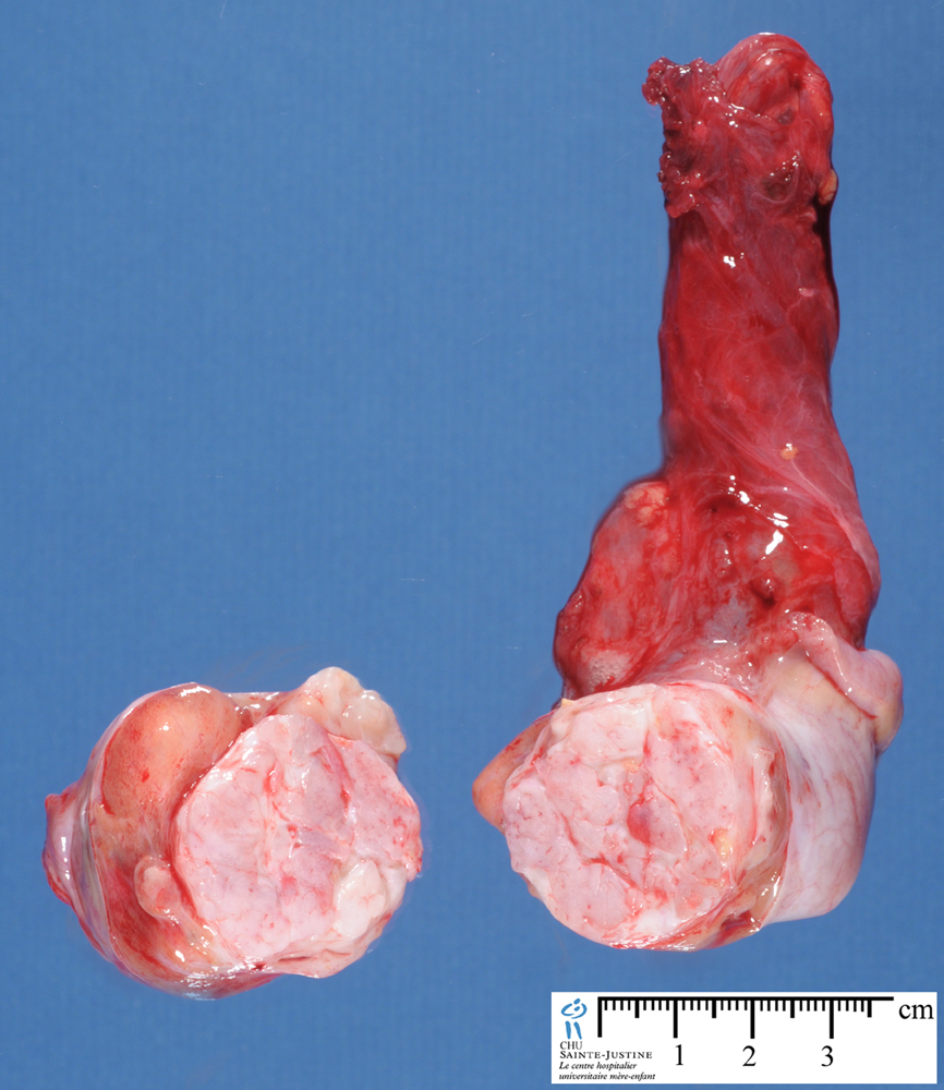
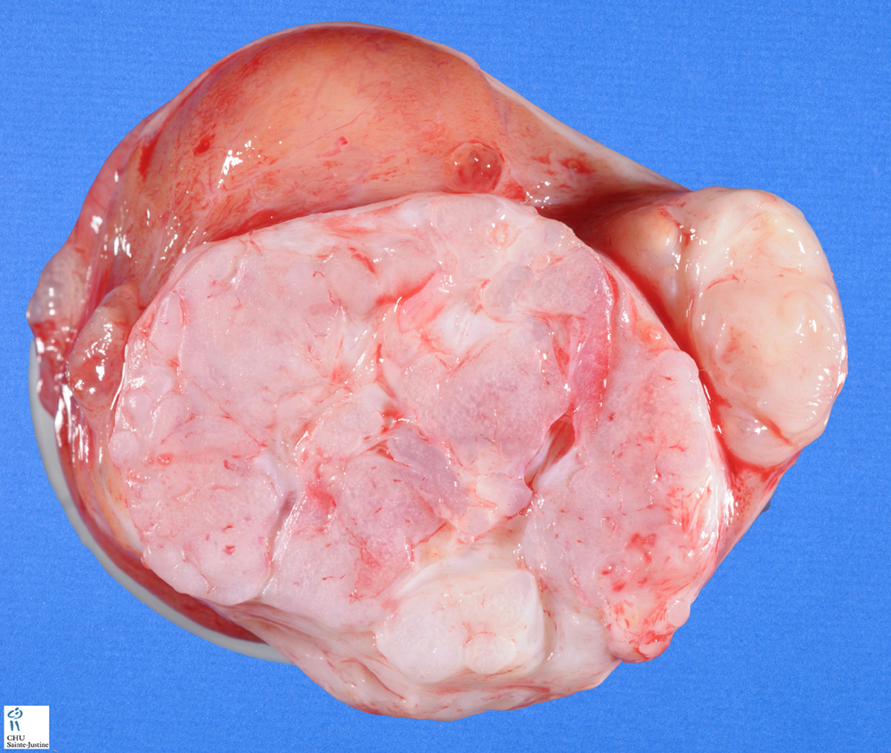
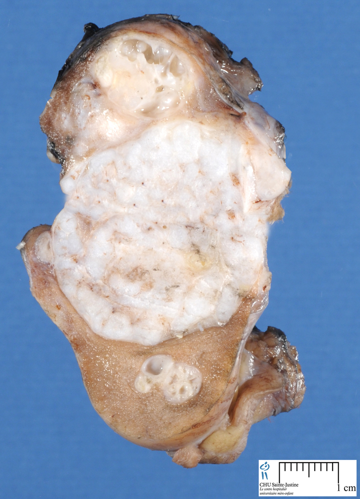
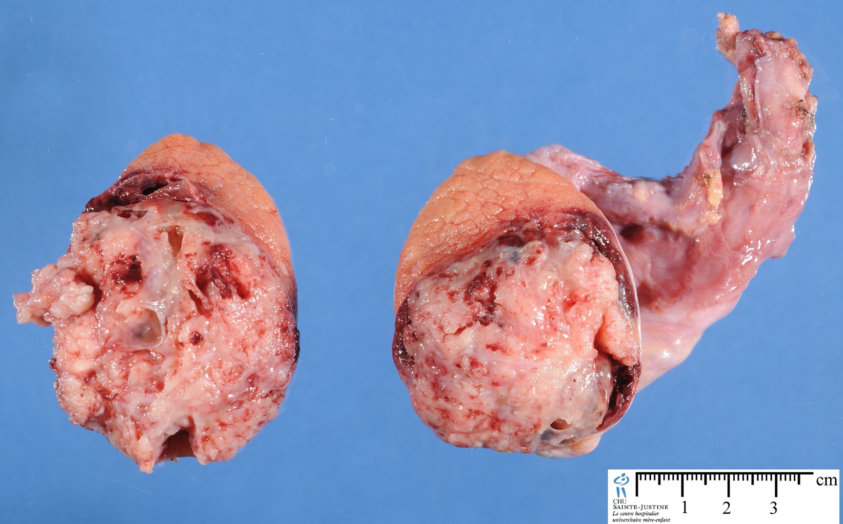
Mixed germ cell tumord are much more common in the testis (33 vs <1%).
Almost any admixture is seen in the testis, many with roughly equal frequency, whereas in the ovary various combinations of yolk sac tumor, dysgerminoma, and teratoma account for the great majority.
Finally, in the testis a random admixture of elements is typical, a more zonal distribution of two or more elements being more usual in the ovary, without implying that the opposite cannot be seen on occasion.
Localization
![]() gonadal ovarian mixed germ cell tumor
gonadal ovarian mixed germ cell tumor
- ovarian mixed germ cell tumor
- -* testicular mixed germ cell tumor