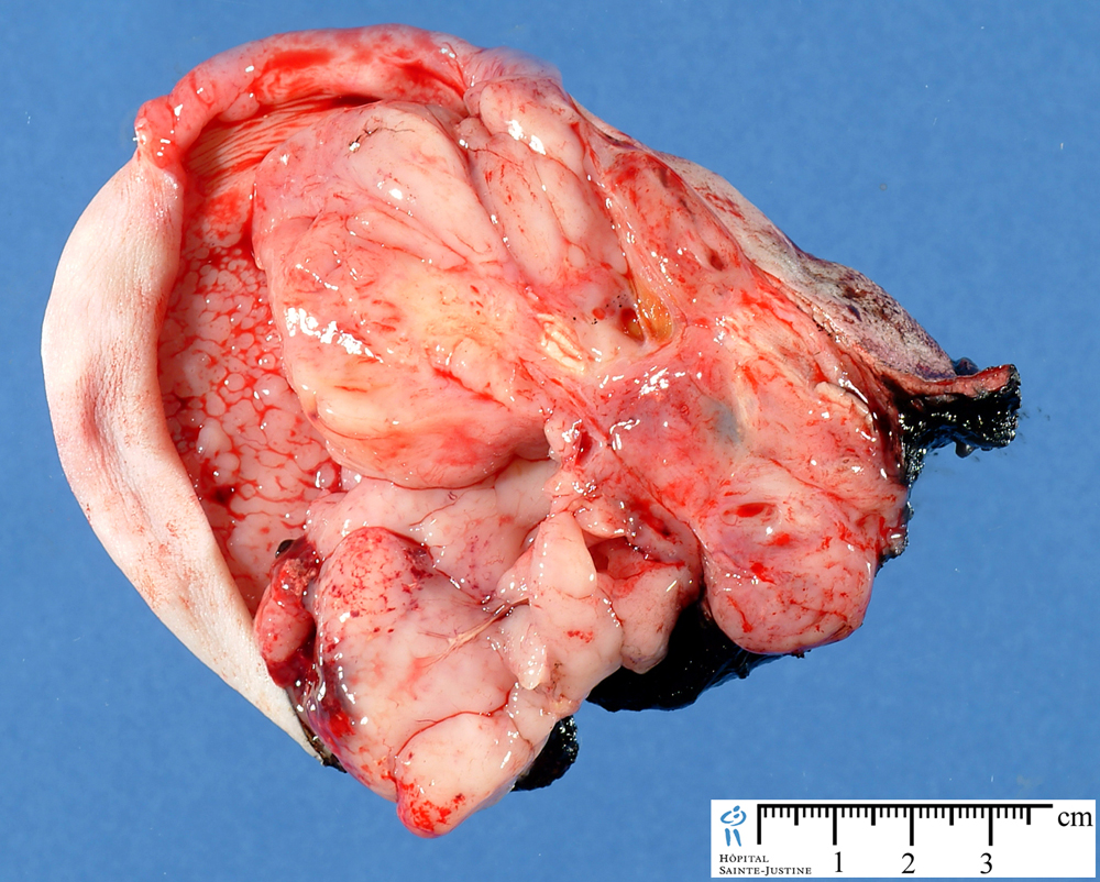
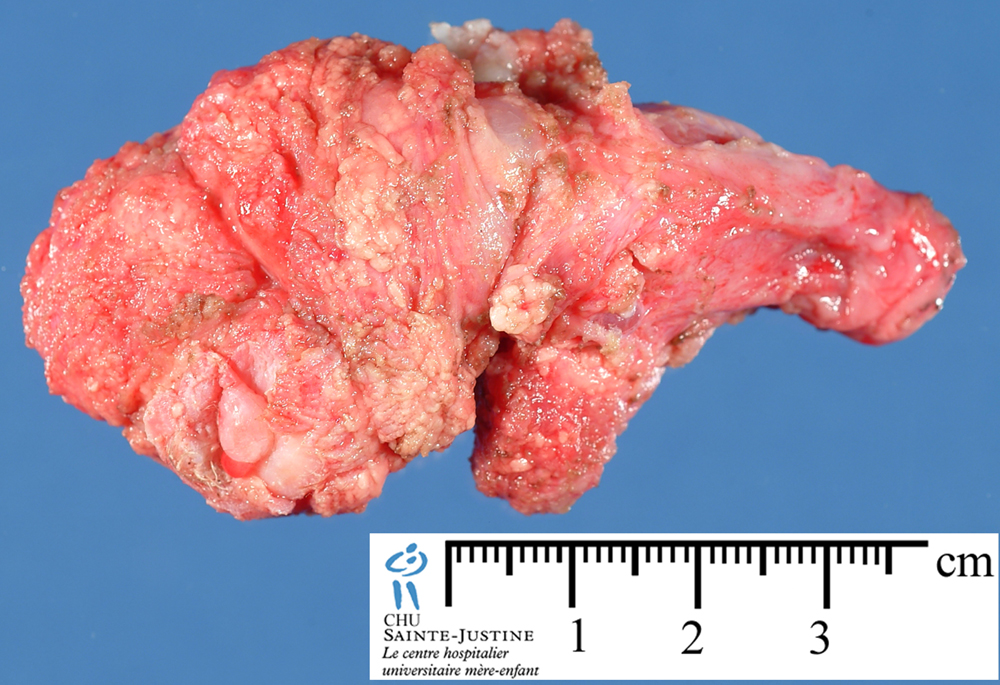
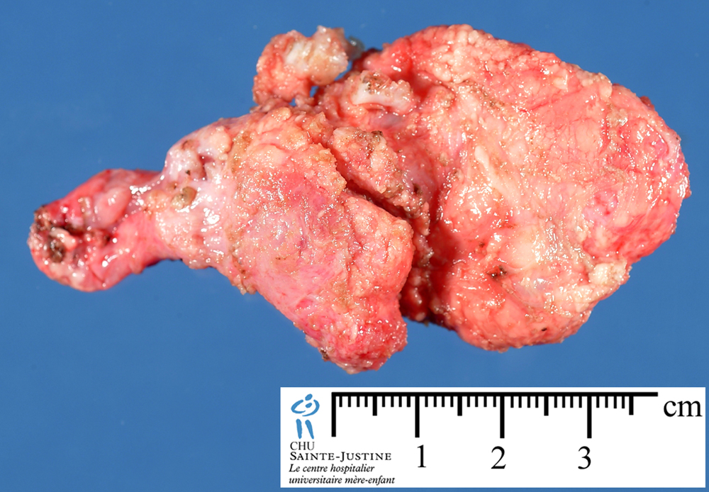
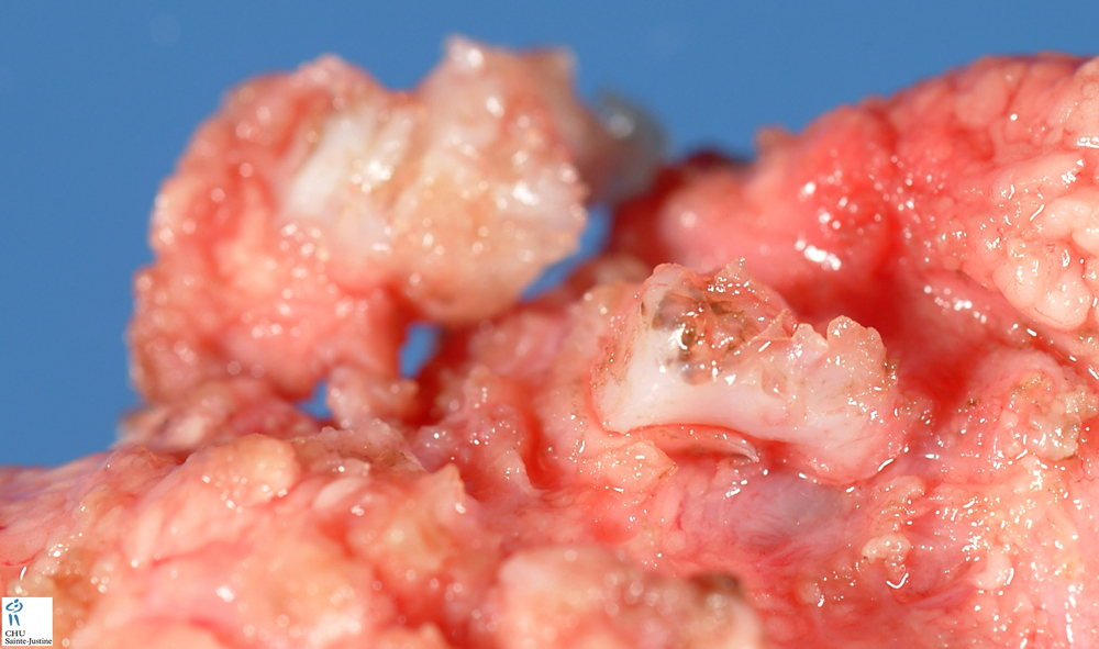
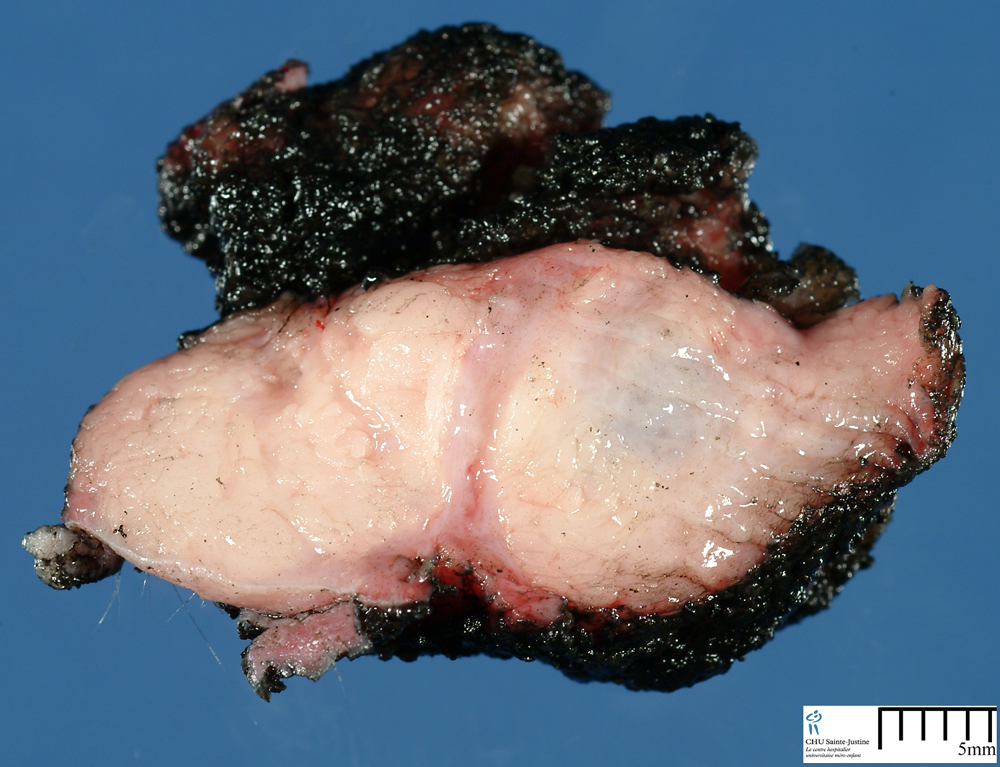
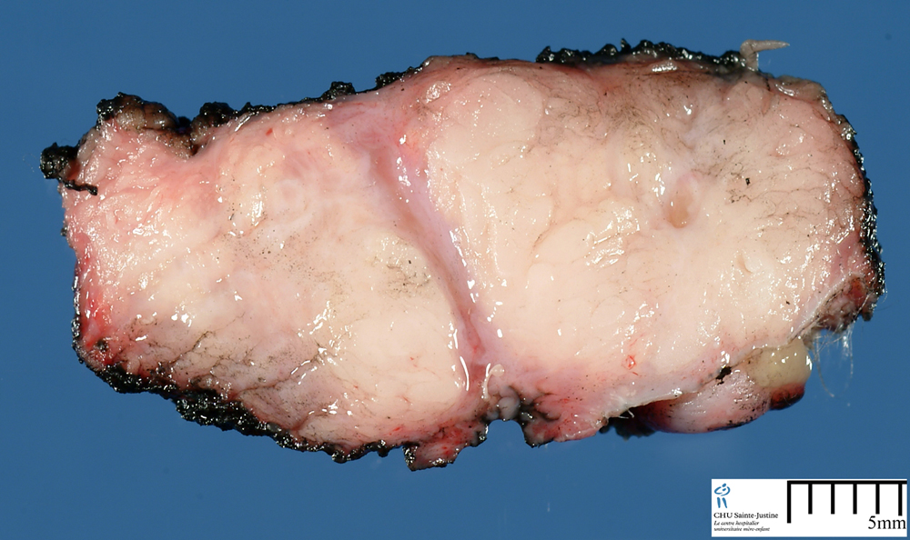
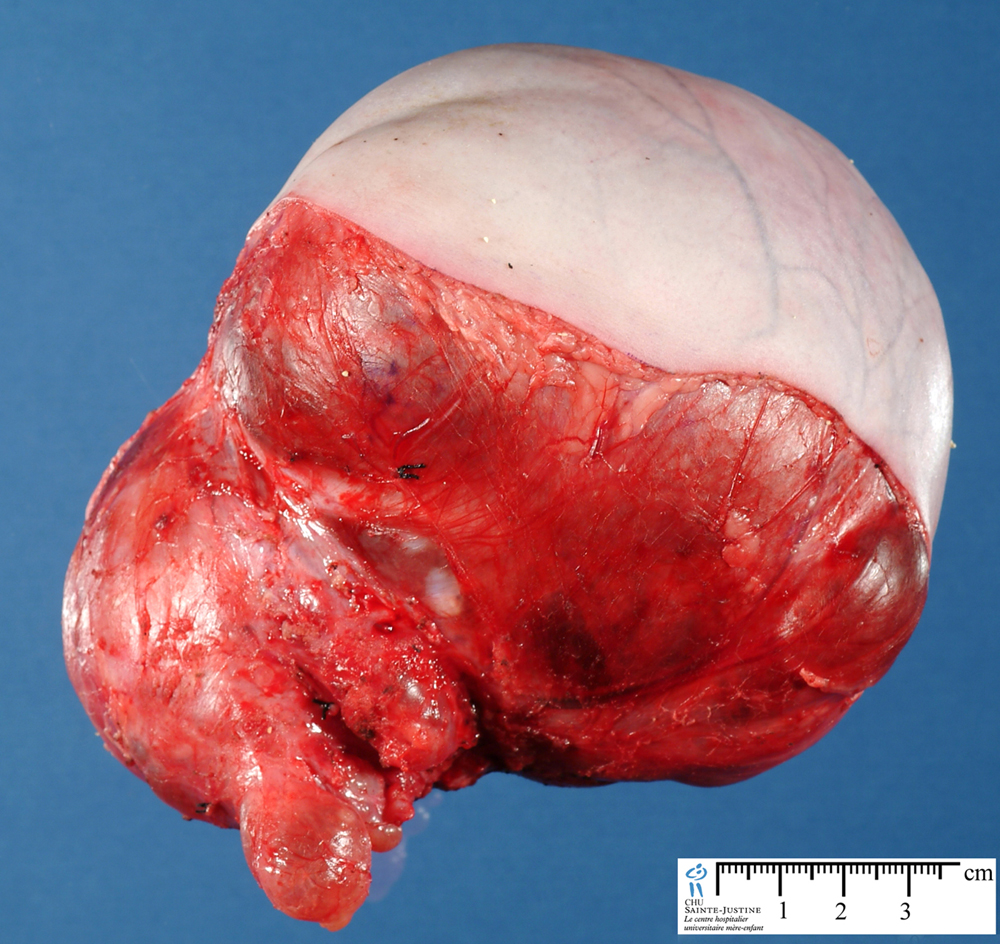
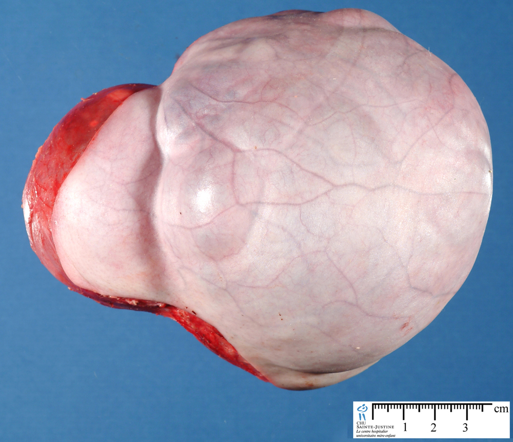
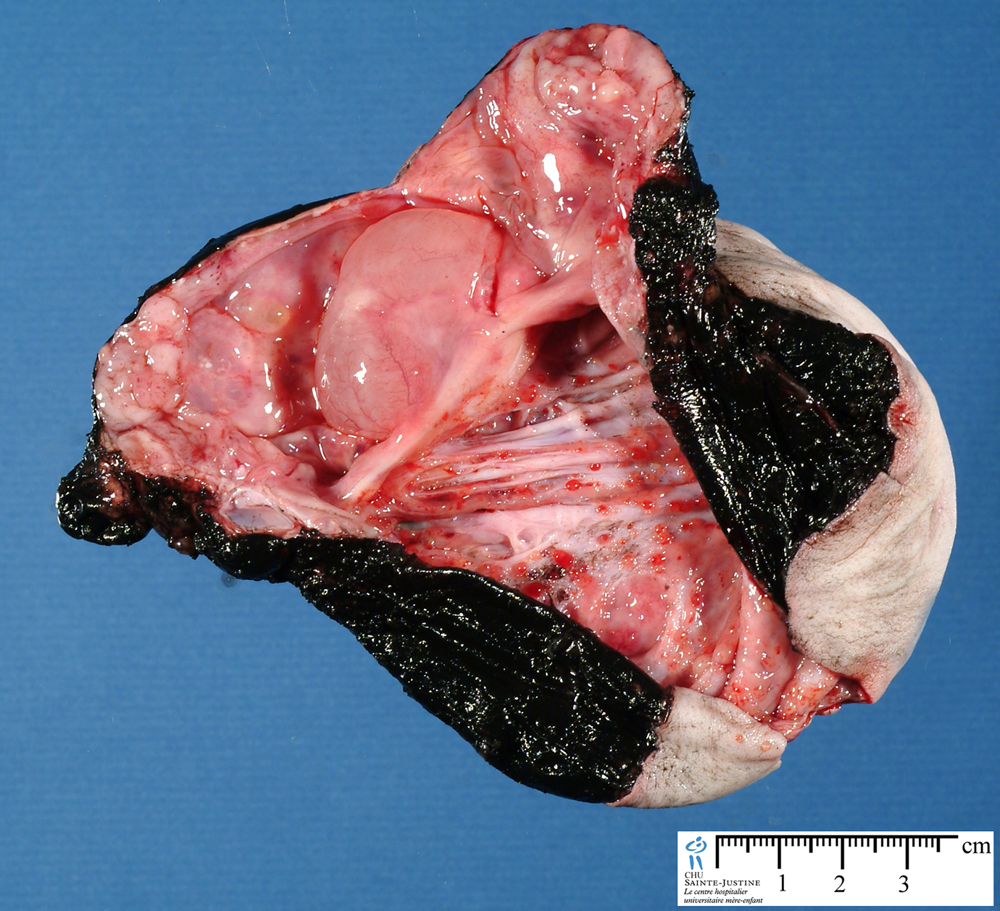
sacrococcygeal teratoma
Image Gallery
[ (||image_reduire{0,60}|inserer_attribut{alt,Sacrococcygeal teratoma}) ] [ (||image_reduire{0,60}|inserer_attribut{alt,Sacrococcygeal teratoma}) ] [ (||image_reduire{0,60}|inserer_attribut{alt,Sacrococcygeal teratoma (Macroscopy)}) ] [ (||image_reduire{0,60}|inserer_attribut{alt,Sacrococcygeal teratoma (SCT) (Macroscopy)}) ] [ (||image_reduire{0,60}|inserer_attribut{alt,Sacrococcygeal teratoma (SCT) (Macroscopy)}) ] [ (||image_reduire{0,60}|inserer_attribut{alt,Sacrococcygeal teratoma (SCT) (Macroscopy)}) ] [ (||image_reduire{0,60}|inserer_attribut{alt,}) ] [ (||image_reduire{0,60}|inserer_attribut{alt,Congenital sacrococcygeal immature teratoma}) ] [ (||image_reduire{0,60}|inserer_attribut{alt,Congenital sacrococcygeal immature teratoma}) ] [ (||image_reduire{0,60}|inserer_attribut{alt,Congenital sacrococcygeal immature teratoma}) ] [ (||image_reduire{0,60}|inserer_attribut{alt,Congenital sacrococcygeal immature teratoma}) ]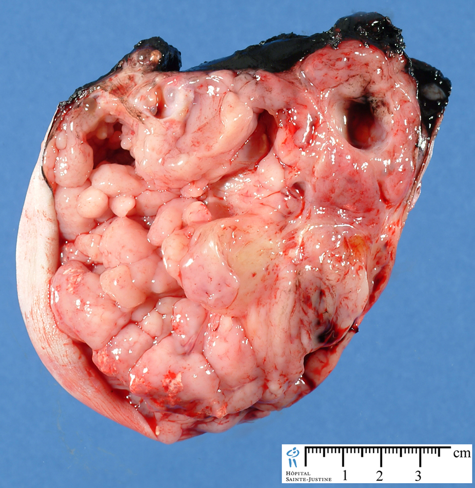
Sacrococcygeal teratoma (SCT) is the most common tumor of the newborn with an incidence of 1 in 35,000 to 40,000 live births.
In the newborn, the sacrococcygeal site is located at the base of the tailbone (coccyx), is the most common location of teratomas in newborns.
Variants
![]() malignant transformation
malignant transformation
- amplification at 8q and 12p (#20113846#)
Cytogenetics
![]() constitutional 7q deletion (#14663834#)
constitutional 7q deletion (#14663834#)
![]() constitutional trisomy 2p (#14663834#)
constitutional trisomy 2p (#14663834#)
![]() constitutional t(12;15)(q13;q25) pat (#12165446#)
constitutional t(12;15)(q13;q25) pat (#12165446#)
- MESDC2/SENP1 fusion gene (#15917269#)
![]() constitutional partial trisomy 10q (10q24.3—>qter) and partial monosomy 17p (p13.3—>pter) (#17295347#)
constitutional partial trisomy 10q (10q24.3—>qter) and partial monosomy 17p (p13.3—>pter) (#17295347#)
Predisposition
![]() Currarino syndrome
Currarino syndrome
- presacral teratoma
- anterior meningocele
- sacral agenesis
- anorectal malformation
See also
![]() congenital germ cell tumors (CGCTs) or neonatal germ cell tumors (NGCTs)
congenital germ cell tumors (CGCTs) or neonatal germ cell tumors (NGCTs)
![]() malignant transformation of sacrococcygeal teratoma (#20113846#)
malignant transformation of sacrococcygeal teratoma (#20113846#)
References
![]() Malignant transformation of an untreated congenital sacrococcygeal teratoma: a amplification at 8q and 12p detected by comparative genomic hybridization. Golas MM, Gunawan B, Raab BW, Füzesi L, Lange B. Cancer Genet Cytogenet. 2010 Feb;197(1):95-8. PMID: #20113846#
Malignant transformation of an untreated congenital sacrococcygeal teratoma: a amplification at 8q and 12p detected by comparative genomic hybridization. Golas MM, Gunawan B, Raab BW, Füzesi L, Lange B. Cancer Genet Cytogenet. 2010 Feb;197(1):95-8. PMID: #20113846#
![]() Immunohistochemical localization of nanog and Oct4 in stem cell compartments of human sacrococcygeal teratomas. Drut R. Histopathology. 2009 May;54(6):763; PMID: #19438750#
Immunohistochemical localization of nanog and Oct4 in stem cell compartments of human sacrococcygeal teratomas. Drut R. Histopathology. 2009 May;54(6):763; PMID: #19438750#
![]() Immunohistochemical localization of nanog and Oct4 in stem cell compartments of human sacrococcygeal teratomas. Busch C, Oppitz M, Wehrmann M, Schweizer P, Drews U. Histopathology. 2008 May;52(6):717-30.PMID: #18439155#
Immunohistochemical localization of nanog and Oct4 in stem cell compartments of human sacrococcygeal teratomas. Busch C, Oppitz M, Wehrmann M, Schweizer P, Drews U. Histopathology. 2008 May;52(6):717-30.PMID: #18439155#
![]() Heerema-McKenney A, Harrison MR, Bratton B, Farrell J, Zaloudek C. Congenital teratoma: a clinicopathologic study of 22 fetal and neonatal tumors. Am J Surg Pathol. 2005 Jan;29(1):29-38. PMID: #15613854#
Heerema-McKenney A, Harrison MR, Bratton B, Farrell J, Zaloudek C. Congenital teratoma: a clinicopathologic study of 22 fetal and neonatal tumors. Am J Surg Pathol. 2005 Jan;29(1):29-38. PMID: #15613854#
![]() Sebire NJ, Fowler D, Ramsay AD. Sacrococcygeal tumors in infancy and childhood; a retrospective histopathological review of 85 cases. Fetal Pediatr Pathol. 2004 Sep-Dec;23(5-6):295-303. PMID: #16137166#
Sebire NJ, Fowler D, Ramsay AD. Sacrococcygeal tumors in infancy and childhood; a retrospective histopathological review of 85 cases. Fetal Pediatr Pathol. 2004 Sep-Dec;23(5-6):295-303. PMID: #16137166#
![]() Prenatal diagnosis of sacrococcygeal teratoma with constitutional partial monosomy 7q/trisomy 2p. Le Caignec C, Winer N, Boceno M, Delnatte C, Podevin G, Liet JM, Quere MP, Joubert M, Rival JM. Prenat Diagn. 2003 Dec 15;23(12):981-4. PMID: #14663834#
Prenatal diagnosis of sacrococcygeal teratoma with constitutional partial monosomy 7q/trisomy 2p. Le Caignec C, Winer N, Boceno M, Delnatte C, Podevin G, Liet JM, Quere MP, Joubert M, Rival JM. Prenat Diagn. 2003 Dec 15;23(12):981-4. PMID: #14663834#
![]() Heifetz SA, Cushing B, Giller R, Shuster JJ, Stolar CJ, Vinocur CD, Hawkins EP. Immature teratomas in children: pathologic considerations: a report from the combined Pediatric Oncology Group/Children’s Cancer Group. Am J Surg Pathol. 1998 Sep;22(9):1115-24. PMID: #9737245#
Heifetz SA, Cushing B, Giller R, Shuster JJ, Stolar CJ, Vinocur CD, Hawkins EP. Immature teratomas in children: pathologic considerations: a report from the combined Pediatric Oncology Group/Children’s Cancer Group. Am J Surg Pathol. 1998 Sep;22(9):1115-24. PMID: #9737245#
![]() Sacrococcygeal teratoma in a fetus with prenatally diagnosed partial trisomy 10q (10q24.3—>qter) and partial monosomy 17p (p13.3—>pter). Batukan C, Ozgun MT, Basbug M, Caglayan O, Dundar M, Murat N. Prenat Diagn. 2007 Apr;27(4):365-8. PMID: #17295347#
Sacrococcygeal teratoma in a fetus with prenatally diagnosed partial trisomy 10q (10q24.3—>qter) and partial monosomy 17p (p13.3—>pter). Batukan C, Ozgun MT, Basbug M, Caglayan O, Dundar M, Murat N. Prenat Diagn. 2007 Apr;27(4):365-8. PMID: #17295347#