PRNP
MIM.176640
Image Gallery
[ (||image_reduire{0,60}|inserer_attribut{alt,Prion protein structure}) ]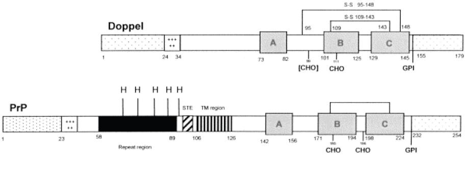
Primary Structure
An analysis of the primary structure of PrP gives insight into its function.
As aforementioned, PrP binds Cu 2+ in order to mediate copper ion recycling within the cell.
It was previously thought that PrP protein bound 4 copper molecules with increasing affinities to its octarepeat region ( PHGGGWGQ).
Copper binds more tightly to the PrP(91-115) domain than the octarepeat region. More specifically, they found that Cu 2+ ions bound to HIS 96 and HIS 111 regions within the PrP(91-115) domain. This domain may act as a copper dependent hinge whereby copper first binds to HIS regions which then activate the octarepeat regions to bind and transport copper as needed.
The PrP(91-115) domain, with HIS 96 and 111 regions, is thought to be the domain that misfolds to yield the deadly PrP Sc conformation of the prion protein.
Secondary Structure
PrP C, the conformation which occurs in healthy, non-infected cells, is composed of 3 alpha helices and 2 beta-pleated sheets. Although the structure of PrP Sc has not yet been ascertained, it is predicted that it is composed of primarily beta pleated sheets as opposed to alpha helices.
The PrP C conformation is composed of approximately 40% small alpha, Greek-helical content with little or no small beta, Greek sheet content, while the PrP 27–30 form contains approximately 50% small beta, Greeksheet and only 20% small alpha, Greek-helical. It is thought that one of the alpha helices in PrP C is misfolded and converted to a beta sheet in PrP Sc.
Of the three alpha helices of PrP C, two of them are longer than the third, indicating that this third is intrinsically less stable due to the decreased number of hydrogen bonds in the coil. Consequently, it is thought that this more unstable, shorter alpha helix, may be that which is converted to the beta sheet.
Between two of the alpha helices of PrP C, there is evidence of a single cystine bond (cys- S-S- cys). These bonds are typical of secreted proteins and those located near the plasma membrane, as is PrP. This cystine bond is thought to stabilize the two alpha helices that it spans in PrP C.
However, in the PrP Sc conformation, this cystine bonds is though to be broken, destabilizing the protein and exposing the unbound sulfurs on the cystine complex towards the outside of the protein. This loss of the cystine bond is thought to be a consequence of 1) a mutation in the gene encoding normal PrP C, 2) random misfolding of a PrP C protein or 3) a result of the interaction of PrP C with PrP Sc , among others.
Functions of the PrP proteins
There is still some debate regarding their exact function, however it is known that the protein is expressed primarily in the central nervous system (CNS).
![]() anti-apoptotic
anti-apoptotic
![]() anti-oxidant: functions as SOD1, sequesters metals (Cu).
anti-oxidant: functions as SOD1, sequesters metals (Cu).
![]() synaptic
synaptic
![]() regulation of sleep
regulation of sleep
![]() signal Transduction ?
signal Transduction ?
![]() myelination
myelination
Cellular prion protein, PrP, provides neuroprotection through copper homeostasis, antioxidant activities, and the inhibition of cell apoptosis.
PrP is a glycoprotein tethered to the cell surface via a glycosyl phosphatidyl inositol (GPI) anchor at the protein’s C terminus.
The presence of free floating PrP in the cytosol indicates that it may also have intracellular functions.
Cu2+ recycling
Prion proteins, or PrPs, are thought to mediate copper ion (Cu 2+) recycling from synaptic clefts, thereby preventing oxidative damage to neurons due to excess Cu 2+.
Studies indicate that PrP C plays a role in copper homeostasis within the cell as elevated synaptic Cu2+ levels are shown to promote PrP C mediated Cu2+ endocytosis.
PrPs control Cu 2+ concentrations within lymphoid or nerve cells by binding extracellular and intracellular Cu2+ ions in their octarepeat regions.
Antioxydant role
PrP C expression in cells was also found to increase antioxidant enzyme activities.
![]() PrP binds copper through octapeptide repeat
PrP binds copper through octapeptide repeat
![]() Interaction with copper changes the conformation of PrP
Interaction with copper changes the conformation of PrP
![]() PrP Null cells are more susceptible to oxidative stress.
PrP Null cells are more susceptible to oxidative stress.
![]() When bound to Copper, PrP acts like superoxide
dismutase.
When bound to Copper, PrP acts like superoxide
dismutase.
![]() PrP Infections increase oxidative stress in brain : increases lipid peroxidation, decreases SOD and Gluthathione reductase activity.
PrP Infections increase oxidative stress in brain : increases lipid peroxidation, decreases SOD and Gluthathione reductase activity.
PrP and apoptosis
PrP is also thought to prevent apoptosis through inhibition of the mitochondrial apoptotic pathway.
The role of PrP in cell survival was suggested by a conserved sequence homology between the BH2 domain of the Bcl-2 protein (BCL2) and the octarepeat region of PrP.
Bcl-2 protein suppresses cell death by homodimerizing with Bax protein, thereby inhibiting Bax function (BAX). Bax protein induces cell death through the apoptotic pathway by permeating, and thus destroying, the mitochondrial membrane.
Coexpression of both cystolic and membrane-bound PrP with Bax abolished Bax-mediated cell death. They were led to conclude that PrP, like Bcl-2, was involved in antiapoptotic activity.
Neuroprotection
![]() Protects against Doppel-mediated neurodegeneration.
Protects against Doppel-mediated neurodegeneration.
![]() Protects against a mouse ataxic disease caused by N-terminal truncation of the PrP molecule
Protects against a mouse ataxic disease caused by N-terminal truncation of the PrP molecule
![]() Protects against ischemia
Protects against ischemia
![]() Protects against kainic acid induced seizures
Protects against kainic acid induced seizures
Synaptic function
![]() PrP present in pre- and post-synaptic structures.
PrP present in pre- and post-synaptic structures.
![]() Regulates level of Cu++ in synapses.
Regulates level of Cu++ in synapses.
![]() Altered inhibitory GABA-A action and decrease LTP in hippocampal slices of PrP-/- mice.
Altered inhibitory GABA-A action and decrease LTP in hippocampal slices of PrP-/- mice.
![]() Decreased hyperpolarization in null mice.
Decreased hyperpolarization in null mice.
Signal transduction
![]() PrP being a membrane protein, it is logical to assume that it may
transduce certain signals to the cell.
PrP being a membrane protein, it is logical to assume that it may
transduce certain signals to the cell.
![]() Thought to regulate [Ca++]i since the « disease form » disregulates
[Ca++]i
Thought to regulate [Ca++]i since the « disease form » disregulates
[Ca++]i
- in neurites (PrP-/-)
- in synaptosomes
 Activates Fyn (tyrosine kinase)
 - Through antibody crosskinking
Binds p66 ligand and stimulates survival pathways.
Proliferation
![]() Activated lymphocytes : mitogenicity is blocked by anti-PrP.
Activated lymphocytes : mitogenicity is blocked by anti-PrP.
![]() Astrocytes - with 106-126 fragment.
Astrocytes - with 106-126 fragment.
PrP-DNA interaction
![]() Binds DNA in vitro.
Binds DNA in vitro.
![]() Associates with retrovirus ie AIDS
Associates with retrovirus ie AIDS
![]() Resembles NCp7 - a nucleoprotein that forms large complexes with DNA
Resembles NCp7 - a nucleoprotein that forms large complexes with DNA
![]() Binding occurs through the octapeptide repeat region.
Binding occurs through the octapeptide repeat region.
Neuritogenesis
![]() PrP binds to :
PrP binds to :
- laminin, a protein important for neuronal differentiation and migration.
- dystroglycan in rafts
- Precursor to laminin receptor
- Tubulin
- Synaptophysin
Prion Proteins: Structure and Location of PrP
The gene that encodes PrP is located on human chromosome 20. The transcription length of the gene is 855 bp long and includes 2 exons. Translation of the gene encoding PrP yields a protein with 217 residues, or amino acids, and an overall charge of 11.
Once transcribed, the PrP protein can adopt one of 2 conformations. Both conformations, PrP C and PrP Sc , have the same primary structure, or amino acid sequence, but differ in their secondary and tertiary structures.
Pathology
| Prion Normal | Pathological Prion |
| Alpha helical structure | Beta sheet |
| Sensitive to Proteases | Resists proteases |
| Cell surface localization mostly, 10% in cytosol | Lysosomal localization |
| Soluble | Insoluble and sometimes forms an amyloid |
| Neuroprotective | Induces brain disease |
An altered form of the normal prion protein, known as PrP Sc (for PrP scrapie), has been found to cause fatal spongiform encephalopathies (SE) including Creutzfeldt-Jakob disease, mad cow disease and scrapie.
These faulty PrP Sc prions convert normal PrP C prions into more PrP Sc , making them a class of unique infectious agents that multiply without a nucleic acid genome. Hence, these proteins were named prions meaning “protenaceous infectious particles”.
Prion diseases: An Overview
The process by which PrP Sc causes spongiform encephalopathies can be summarized in four steps:
![]() PrP Sc enters or is created as a result of mutation in non-infected cells.
PrP Sc enters or is created as a result of mutation in non-infected cells.
![]() PrP Sc converts PrP C to more PrP Sc.
PrP Sc converts PrP C to more PrP Sc.
![]() Protective proteinases within the cell attack and truncate PrP Sc.
Protective proteinases within the cell attack and truncate PrP Sc.
![]() After conversion and truncation, PrP Sc (27-30) form aggregations within the cell.
After conversion and truncation, PrP Sc (27-30) form aggregations within the cell.
Prion infections
Faulty PrP Sc conformations may enter the body through consumption of scrapie-infected cells or are produced as a consequence of mutations in the gene encoding normal PrP C.
PrP Sc can also enter the body through the consumption of meat of cows infected with mad cow disease or sheep infected with scrapie.
PrP mutations
In humans, mutations within the Human PrP gene cause Creutzfeldt-Jakob disease (CJD), Gerstmann-Straussler-Scheinker disease, and Fatal Fetal Insomnia.
Conversion of PrP C to PrP Sc
The conversion of PrP C to PrP Sc is thought to be mediated by direct interactions between PrP C and PrP Sc . This is evidenced by mice PrP-/- knockout studies yielding mice resistant to scrapie infection.
PrP-/- knockout mice were viable because the role of PrP is so important that its function in neuroprotection is redundant. In support of these findings, studies of chaperone mediated PrP to PrP Sc conversion demonstrate that while some chaperones could induce transformation of PrP C, none could promote conversion in the complete absence of PrP Sc.
Two chaperones thought to mediate this conversion are GroEL and Hsp 104 (heat shock protein 104) (DebBurman et al., 1997).
Attack by Proteinases
PrP Sc (27-30) is created when proteinases within the cell, which are made to destroy foreign proteins, attempt to eradicate PrP Sc through digestion but instead only truncate it.
Indeed protease resistance is one of the primary characteristics of PrP Sc protein and is used as a diagnostic tool for cells that are thought to be scrapie-infected. Whereas normal PrP C is sensitive to proteinase K digestion, PrP Sc loses only 50-80 amino acids in its N terminus and another 70-80 amino acids in its C terminus.
Formation of amyloid plaques
Once created, this protease resistant core, with its compact size and instability, aggregates with other PrP Sc (27-30) fragments to form aggregations called amyloid plaques. Recall that in PrP Sc , a normally intact cystine bond is disrupted, yielding exposed and unstable sulfur atoms that orient themselves outward from the protein.
Exposed sulfurs on PrP Sc (27-30) fragments bind to one another creating growing disulfide polymers within the cell. Ultimately, the cells lyse, releasing more PrP Sc into extracellular spaces much like viruses in their lytic cycles.
Pathology
![]() germline mutations in the prion protein gene PRNP
germline mutations in the prion protein gene PRNP
- Gerstmann-Straussler disease (MIM.137440)
- Creutzfeldt-Jakob disease (MIM.123400)
- familial fatal insomnia (MIM.600072)
![]() aberrant isoforms of the prion protein as an infectious in kuru (MIM.245300) and in scrapie in sheep.
aberrant isoforms of the prion protein as an infectious in kuru (MIM.245300) and in scrapie in sheep.
See also
![]() prion diseases
prion diseases