Home > E. Pathology by systems > Locomotory system > Tendons and tendon sheaths > tendon sheath fibroma
tendon sheath fibroma
Friday 13 June 2003
fibroma of tendon sheath
| PO | StanMed |
Images
https://twitter.com/drqari/status/848145290108559361
https://twitter.com/ARP_Press/status/1119707546737152000
JRC:6487 : Fibroma of tendon sheath.
Definition: Fibroma of the tendon sheath is a rare tumor described as a benign lesion or a tumor-like reactive lesion arising from the synovium of tendon sheath.
Fibroma of the tendon sheath occurs in both adults and children, with 20% of cases occurring in children and adolescents. These lesions most commonly affect the fingers, hand, and wrist, with thumb involvement in as many as 41% of patients.
Deeply attached to the tendon or tendon sheath, these tumors are usually solitary, with a characteristic lobulated external appearance and usually small, 1–2 cm in diameter. The skin overlying a tendon sheath fibroma, unlike infantile digital fibromatosis lesions, is generally not adherent since the dermis is seldom involved.
Although fibromas of the tendon sheath can be confused with giant cell tumors of the tendon sheath, the giant cell tumor occurs in older patients and contains characteristic macrophages and siderophages, which are not present in the tendon sheath fibroma.
Localization
It has been reported mainly in finger and hand tendons as a benign, slowly growing nodule that arises from a synovial sheath.
Only rare cases of localization within a joint capsule have been described: in the knee (posterior joint capsule, posterior cruciate ligament, suprapatellar pouch), in the radioulnar joint, in the temporomandibular joint, and in the shoulder joint.
Macroscopy
fibrous structure
nonrubbery, hard consistency
gray-pearly white
multilobular
solid
no cystic cavities
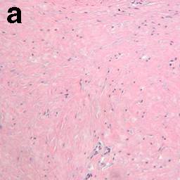
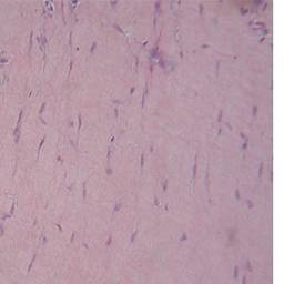
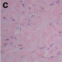
Microscopy
Histologically, fibromas of the tendon sheath are composed of a proliferation of dense fibrous stroma with slit-like vascular channels. Fibroblast spindles are stellate-shaped. Cells may be arranged in fascicles. Other histologic features include myxoid change, osteoid change, hyalinization, and scattered multinucleated giant cells.
variable cellularity
central nodular area composed of dense fibrous connective tissue with focal areas of myxoid degeneration
peripheral dense fibrous connective tissue linked to the tendon sheath with some vascular structures
Fibroma of the tendon sheath, or tenosynovial fibroma, was first defined by Geschickter and Copeland in 1936. It has been described as a fibrotic neoplasm or a reactive fibrosis, but its precise origin is still unclear according to the current classification.
Histologically, it is a poorly recognized, slowly growing, benign proliferation of fibroblasts surrounded by collagen fibers, which appears as a fibrous nodule attached to tendon or tendon sheath; a smooth, dense, multinodular mass with a diffuse pearly white appearance, ranging in size from 0.5 to 5.5 cm.
A dense, matrix-rich collagenous stroma is arranged in nodules with slit-like vascular channels throughout it. Occasionally myxoid and sclerotic regions are seen, depending probably on the vascular impairment due to compression. The cells are mainly spindle shaped and are less frequently stellate.
Many of the cells are represented by myofibroblasts. Seventy-five to eighty-two percent of the tumors have been described in the extremities, most commonly the fingers, hands and wrists.
The most important case report appears to be that of Chung and Enzinger in 1979, who reported on 138 patients: 98% of cases occurred in those locations.
The tumor can occur at any age, with peak incidence occurring between 20 and 50 years. In the same paper, Chung and Enzinger reported a median age of 31. The male:female ratio has been described as 1.5–3:1.
The clinical presentation of tendon sheath fibroma often occurs years after its formation as a painless, slowly growing mass that may irritate the surrounding tissues by compression.
Nerve compression has been described in the distal forearm, presenting itself as a median nerve neuropathy. Less than 10% of patients have reported a history of trauma.
Diagnosis must be based on the patient history and clinical examination, MRI imaging and histology. Plain X-rays are usually negative, except when large masses compress surrounding muscles or fat, or there are erosive bony changes, which are rarely described.
Immunochemistry
vimentin +
muscle-ŝpecific actin +
smooth muscle actin +
desmin -
CD68 +/-
factor XIIIa +
CD34 +
Differential diagnosis
fibrous histiocytoma
nodular fasciitis
giant cell tumor of the tendon sheath
if nuclear pleomorphism: malignant fibrous histiocytoma
Differential diagnosis must be made with giant cell tumor of the tendon sheath (GCTTS), representing a localized manifestation of pigmented villonodular synovitis that is less hyalinized and more cellular, and with histiocytes and monocytes as well as multinucleated giant cells, foam cells and hemosiderin-laden macrophages. Due to similarities between some forms of the two tumors, some authors have hypothesized that they may be two phenotypic extremities of a single entity.
FTS must also be distinguished from nodular fasciitis, which resembles FTS histologically but is a more rapidly growing mass.
Treatment and prognosis
Treatment is by local excision, with a reported recurrence of 24%; all of the cases were described in hands and fingers, and this probably depends on the accuracy of the excision itself. To our knowledge, a malignant transformation has never been described.
Cytogenetics
translocation t(2;11)(q31-32;q12) (2q31-32 and 11q12)
References
Fibroma of tendon sheath located within the ankle joint capsule. Ciatti R, Mariani PP. J Orthop Traumatol. 2009 Sep;10(3):147-50. PMID: 19644650 (Free)
Bertolotto M, Rosemberg I, Parodi RC (1996) Case report: fibroma of tendon sheath in the distal forearm with associated median nerve neuropathy: US, CT and MR appearances. Clin Radiol 51:370–372
Chung EB, Enzinger FM (1979) Fibroma of tendon sheath. Cancer 44:1945–1954
Enzinger FM, Weiss WS (2001) Soft tissue tumors, 4th edn. Mosby, St Louis.
Fox MG, Kransdorf MJ, Bancroft LW (2003) MR imaging of fibroma of the tendon sheath. AJR 180:1449–1453
Geshickter CF, Copeland MM (1949) Tumors of the bone, 3rd edn. JB Lippincott, Philadelphia.
Hashimoto H, Tsuneyoshi M, Daimaru Y, Ushijima M, Enjoji M (1985) Fibroma of tendon sheath: a tumor of myofibroblasts: a clinicopathologic study of 18 cases. Acta Pathol Jpn 35:1099–1107
Hermann G, Hoch BL, Springfield D, Abdelwahab IF, Klein MJ (2006) Intra-articular fibroma of tendon sheath of the shoulder joint: synovial fibroma. Skeletal Radiol 35(8):603–607
Hitora T, Yamamoto T, Akisue T, Marui T, Nagira K, Ohta R, Kurosaka M (2002) Fibroma of thendon sheath originating from the knee joint capsule. Clin Imaging 26(4):280–283
Hur J, Damron TA, Vermont AI, Mathur SC (1999) Fibroma of tendon sheath of the infrapatellar fat pad. Skeletal Radiol 28:407–410
Li TJ, Kitano M, Tsuneyoshi M (1997) Intraarticular fibroma of tendon sheath in the temporomandibular joint. Oral Surg Oral Med Oral Pathol Oral Radiol Endod 84:407–410
Lourie JA, Lwin KY, Woods CG (1992) Case report 734, fibroma of tendon sheath eroding 3rd metatarsal bone. Skeletal Radiol 21:273–275
Misawa A, Okada K, Hirano Y, Sageshima M (1999) Fibroma of tendon sheath arising from the radio-ulnar joint. Pathol Int 49:1089–1092
Ogata K, Ushijima M (1987) Tenosynovial fibroma arising from the posterior cruciate ligament. Clin Orthop Relat Res 215:153–155
Pinar H, Ozkan M, Ozaksoy D, Pabuccuoglu U, Akseki D, Karaoglan O (1995) Intraarticular fibroma of the tendon sheath of the knee. Arthroscopy 11:608–611.
Takakubo Y, Fukushima S, Asano T, Yamakawa M (2005) Intraarticular fibroma of the tendon sheath in the knee. Clin Orthop 439:280–285
Pulitzer DR, Martin PC, Reed RJ (1989) Fibroma of tendon sheath: a clinicopathologic study of 32 cases. Am J Surg Pathol 13:472–479
Smith PS, Pieterse AS, McClure J (1982) Fibroma of tendon sheath. J Clin Pathol 35:842–848
Sundaram M, McGuire MH, Schajowicz F (1981) Soft tissue masses: histologic basis for decreased signal (short T2) on T2-weighted MR images. AJR 148:1241–1250.v