photoreceptors
Image Gallery
[ (||image_reduire{0,60}|inserer_attribut{alt,}) ]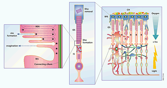
The retina consists of three main cellular layers (right): a rod cells (R) and cone cells (C) layer or photoreceptor layer, a layer of interneurons (bipolar interneurons, horizontal interneurons, amacrine cells) and the ganglion cell layer, whose axons form the optic nerve, connecting retina and brain.
The photoreceptors are oriented in the same plane as incoming light, which funnels through the inner to the OS.
The outer segment contains a dense array of light-sensitive, hollow discs, whose membranes are packed with rhodopsin (in rod cells) or color opsin (cone cells).
Outer segment shortening occurs in several models of retinal degeneration, including Rom1 knockout mice5, which may lead to increased oxidative damage from exposure to high oxygen pressure levels.
The inner and outer segments of photoreceptors are connected by a modified cilium.
Models of disc formation propose an evagination of the ciliary plasma membrane, followed by a zippering process, which fuses adjacent disc-like protrusions at the rim margins, separating them from the plasma membrane.
The length of the outer segment is determined by the relative rates of disc formation at the base and removal by retinal pigment epithelial (RPE) cells underlying the retina.
Pathology
![]() photoreceptor degeneration
photoreceptor degeneration
References
![]() Pacione LR, Szego MJ, Ikeda S, Nishina PM, McInnes RR. Progress toward understanding the genetic and biochemical mechanisms of inherited photoreceptor degenerations. Annu Rev Neurosci. 2003;26:657-700. PMID: #14527271#
Pacione LR, Szego MJ, Ikeda S, Nishina PM, McInnes RR. Progress toward understanding the genetic and biochemical mechanisms of inherited photoreceptor degenerations. Annu Rev Neurosci. 2003;26:657-700. PMID: #14527271#