Home > E. Pathology by systems > Reproductive system > Male genital system > Testis > testicular yolk sac tumor
testicular yolk sac tumor
Monday 8 March 2004
mesonephroma ovarri; endodermal sinus tumor of the testis; yolk sac tumor of the testis
Definition: A malignant germ cell neoplasm that recapitulates the primary embryonic yolk sac tissue.
The tumor was also called ’mesonephroma ovarri’ due to the finding of glomeruli like structure, the Schiller-Duval body. There are actually blood vessels surrounded by primordial germ cells, a finding first noted in rat endoderm.
Epidemiology
Age: six months to five years, peak in boys of age less than 2 years
Incidence: YST is the most common malignant testicular germ cell tumor of pediatric population (70%)
Pathogenesis
Hypermethylation of the RUNX3 gene promoter
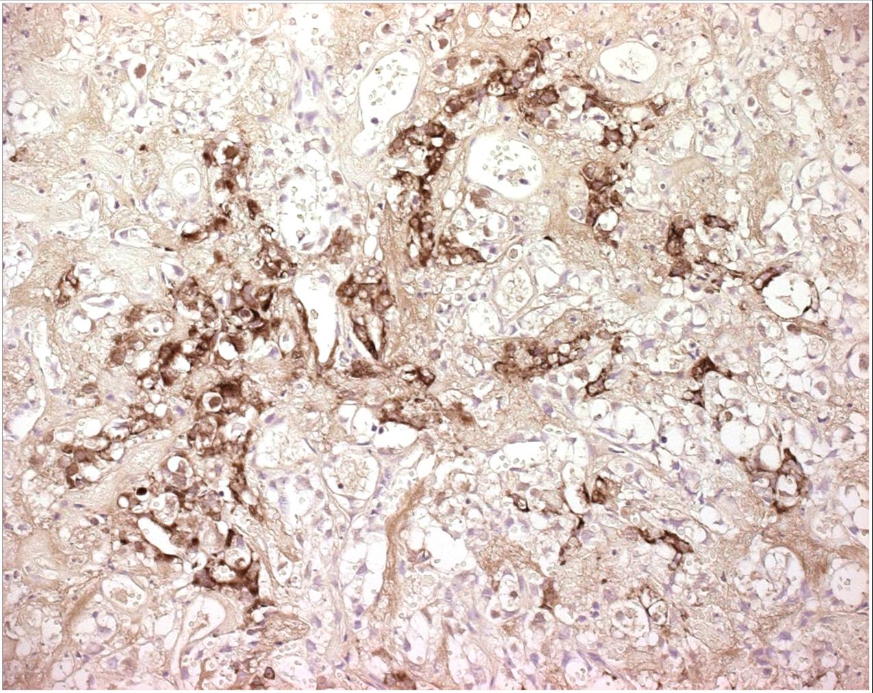
Pediatric cases express GATA-4, a transcription factor that regulates differentiation and function of murine yolk sac endoderm
Treatment
1. Surgical Management · Radical Orchiectomy with high ligation of the spermatic cord · Retroperitoneal Lymph Node Dissection – Indications a. The presence of a persistent retroperitoneal mass following chemotherapy b. Persistent elevation of serum AFP following chemotherapy with no evidence of metastases on imaging studies c. Normal or unknown serum AFP levels at time of diagnosis
2. Chemotherapy Management · Platinum-based regimens seems to apply to childhood germ cell Prognosis Excellent (5 year survival 90% even in advanced cases)
Prepubertal YST
Mostly shows pure histology Isochrome 12p is not seen.
Intratubular neoplasia is absent.
Euploid or tetraploid
Recurrent nonrandom chromosomal abnormalities suggestive of a different pathway for embryogenesis of YST
- 1p deletion
- 3p duplication
- 6q deletion
- p53 gene mutations absent.
Equal incidence of lymphatic and hematogenous spread for metastases.
Presents as stage I disease (85%)
Adult YST
Presents along with other germ cell tumor components
Isochrome 12p positive
Intratubular neoplasia is present
Usually aneuploid
p53 gene mutations
Lymphatic route is primarily spread for metastases
Often presents as higher stage disease
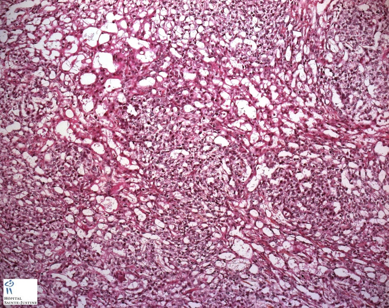
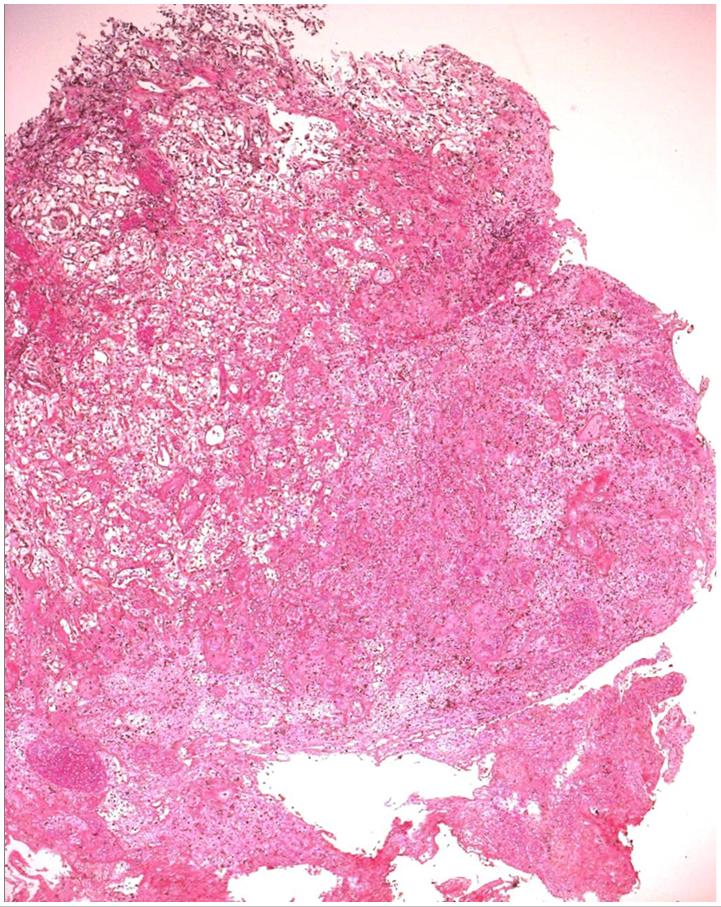
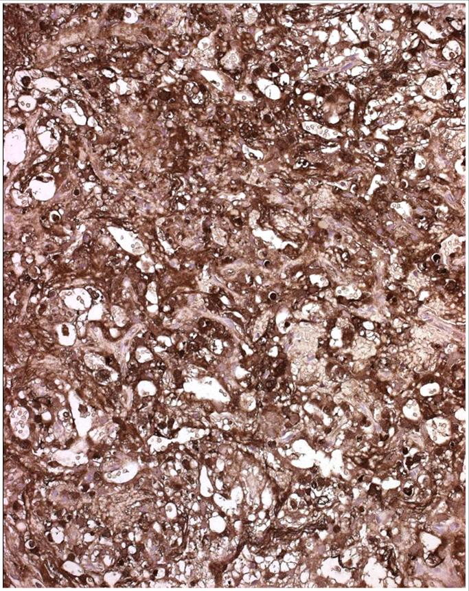
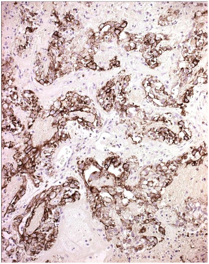
Architectural patterns
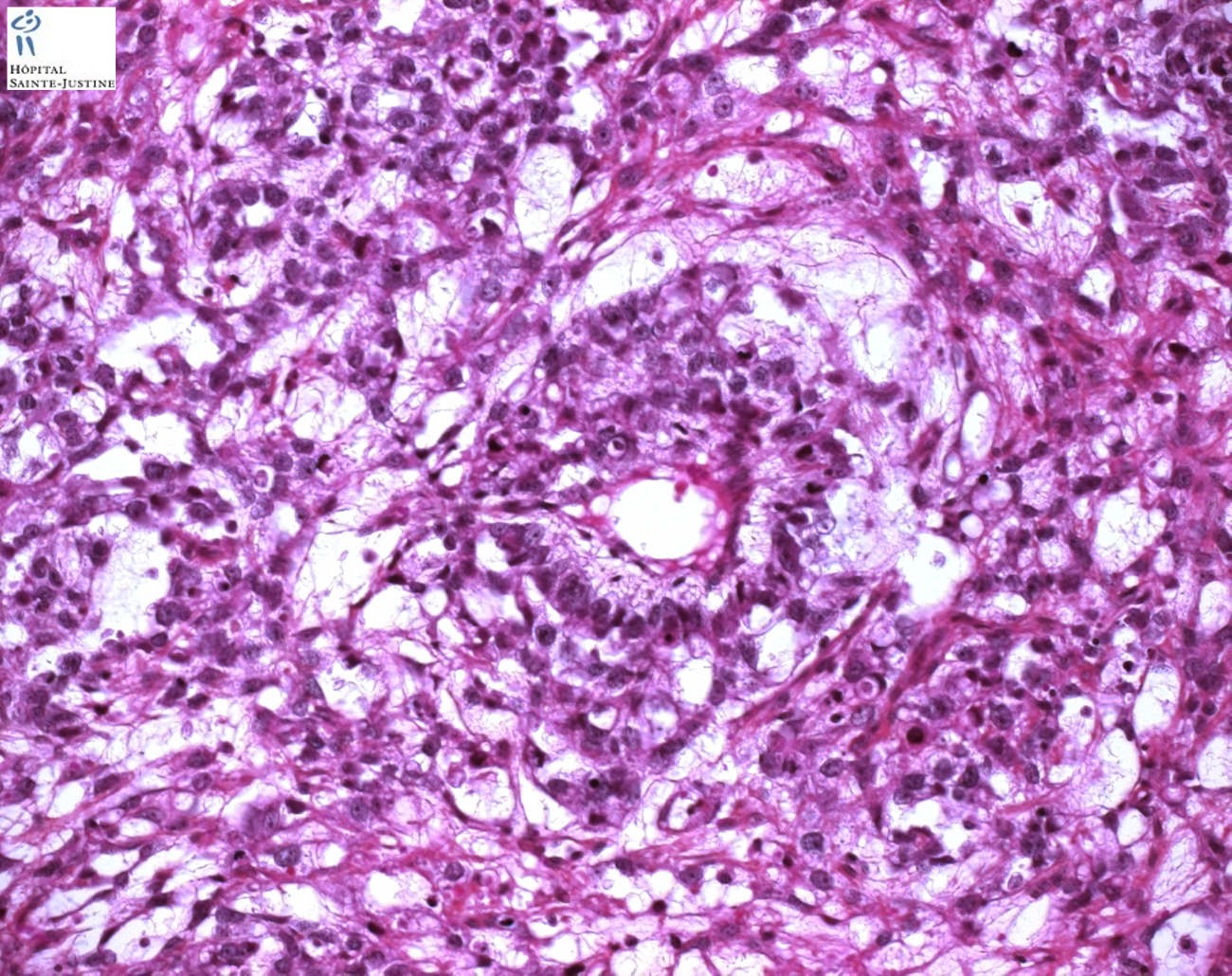
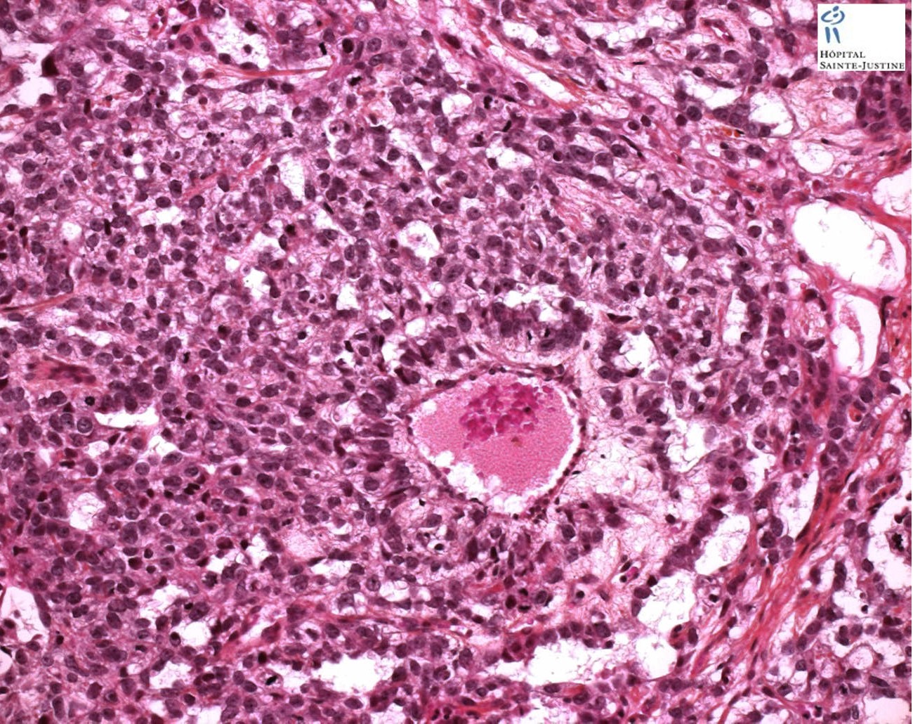
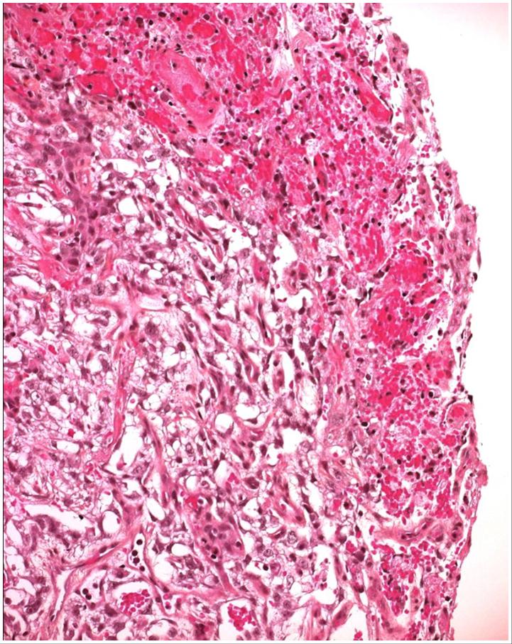
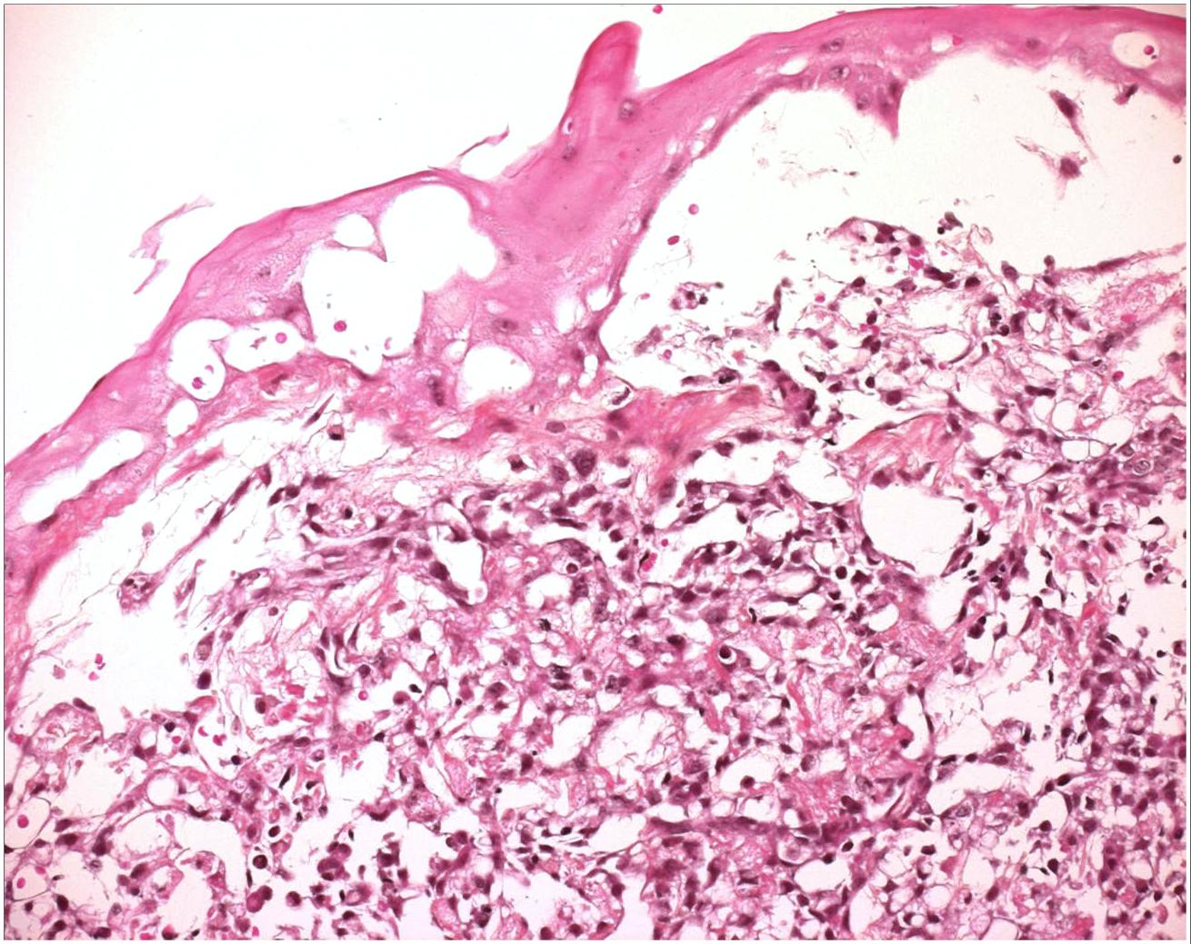
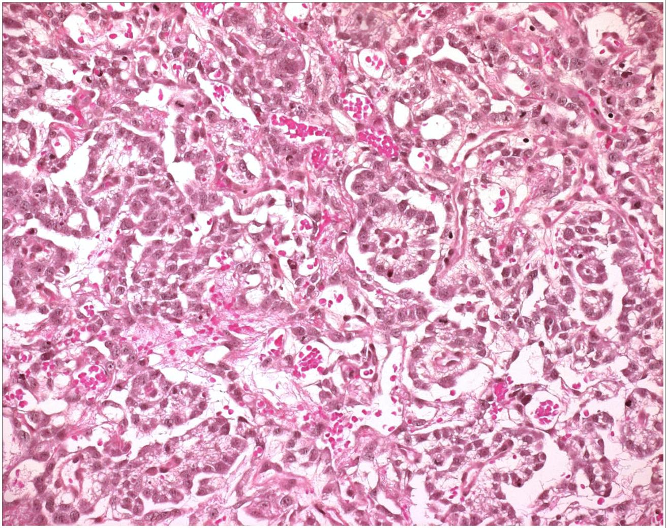
Schiller-Duvall bodies (Schiller-Duvall body).
- Schiller-Duvall body is a structure seen in the endodermal sinus pattern of yolk sac tumor. It consists of a central vessel surrounded by tumor cells – the whole structure being contained in a cystic space often lined by flattened tumor cells.
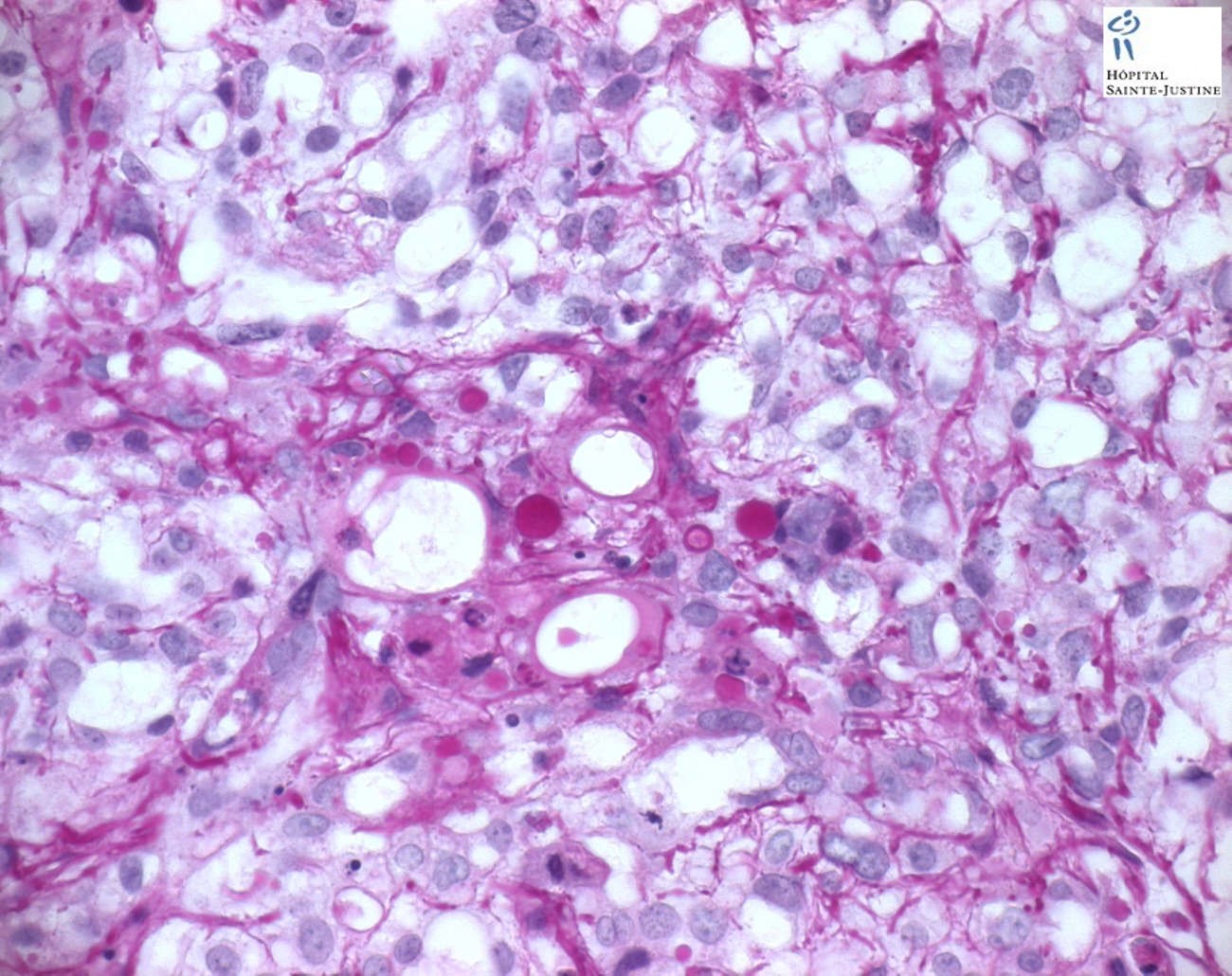
hyaline globules
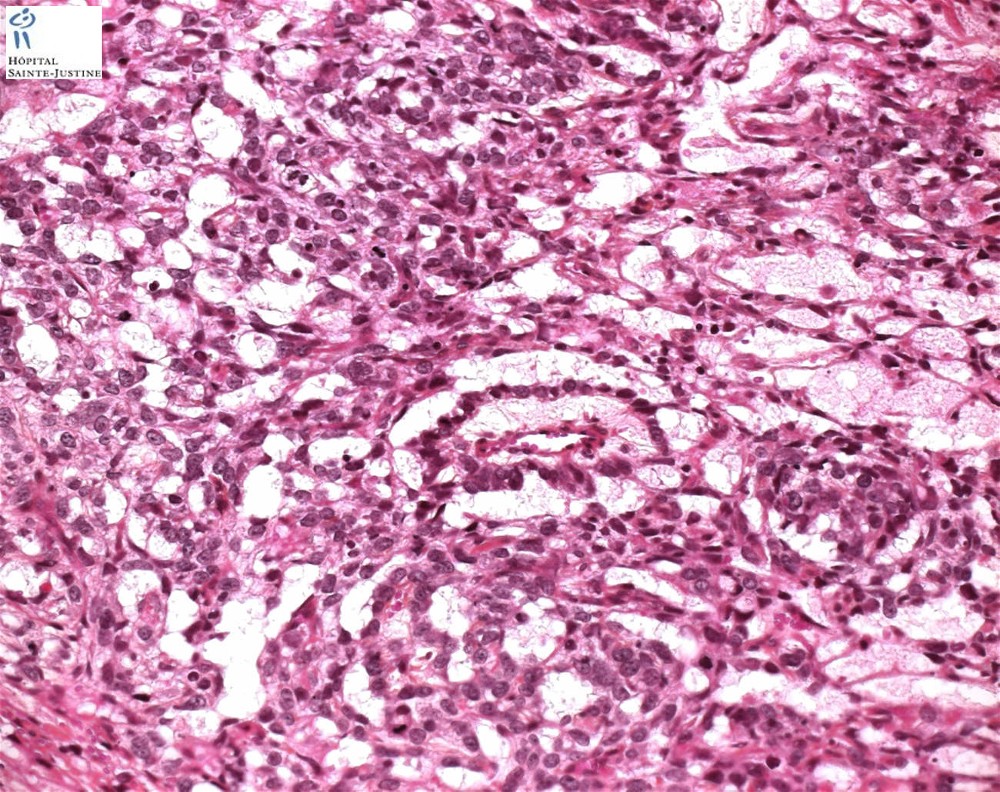
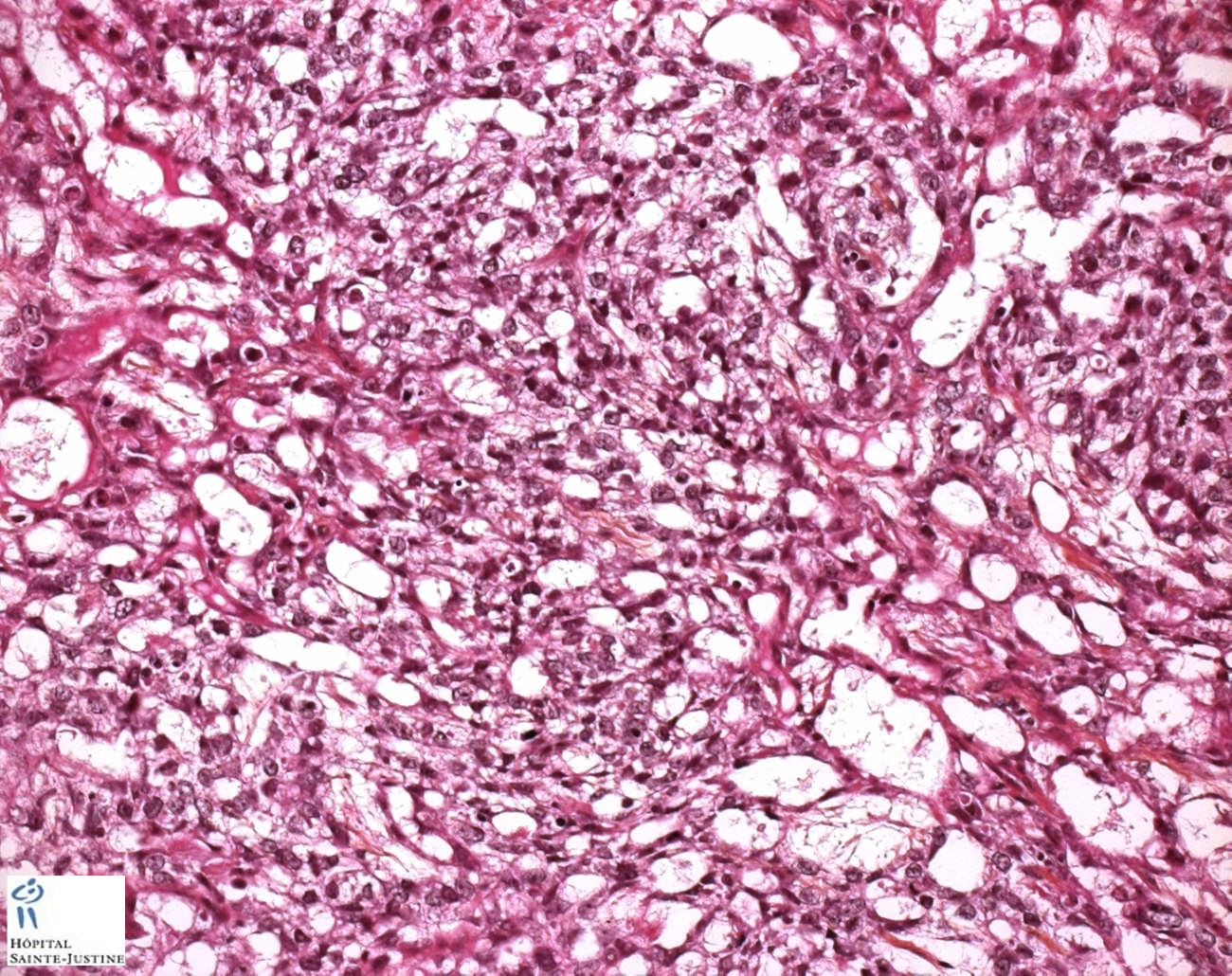
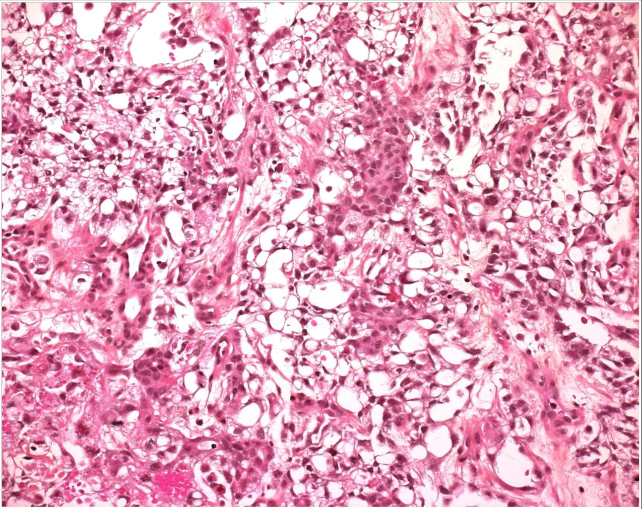
microcystic pattern: interconnecting cords and ribbons of tumor cells are surrounded by abundant myxoid stroma. Intracellular vacuoles and microcystic areas may sometimes resemble lipoblasts; however, the tumor cells do not contain lipid. Microcystic pattern often coexists with other architectural patterns. The tumor cells have vesicular nuclei with punctate nucleoli. Wisps of myxoid material are seen in cystic spaces. This accounts for gelatinous appearance on cut surface of the tumor.
myxomatous pattern: neoplastic stellate, spindle, or epithelioid cells in abundant myxoid stroma. Many of these cells are pluripotential and can form skeletal muscle, cartilage, and bone. Such areas should not be confused with teratoma
hepatoid pattern (20%) (hepatoid yolk sac tumor)
- sheets of polygonal cells with abundant eosinophilic cytoplasm
- hyaline globules
- bile canaculi
solid pattern
- sheets of uniform tumor cells with clear or pale pink cytoplasm
- differential diagnosis: seminoma (It lacks the fibrous septa with lymphoid infiltrate seen in seminoma. )
Other localizations
ovarian yolk sac tumor
vaginal yolk sac tumor
prostatic yolk sac tumor
mediastinal yolk sac tumor
Differential diagnosis (Examples)
microcystic Leydig cell tumors (10328086)
CGH (15880464)
| CGH losses | |
| 1p35-pter | |
| CGH gains | |
| 3p21-pter | 20q13 |
Case records
Case humpath.com #12475: Vaginal yolk sac tumor
pathxchange.org #11530
Web references
References
Yolk Sac Tumor of the Testis in Infants and Children: A Clinicopathologic Analysis of 33 Cases. Cornejo KM, Frazier L, Lee RS, Kozakewich HP, Young RH. Am J Surg Pathol. 2015 Mar 30. PMID: 25828390