congenital pulmonary airway malformation
Image Gallery
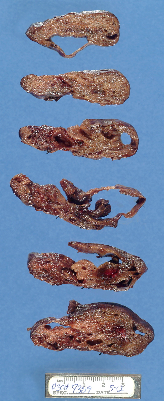
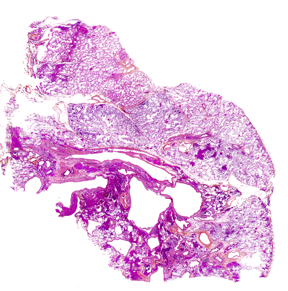
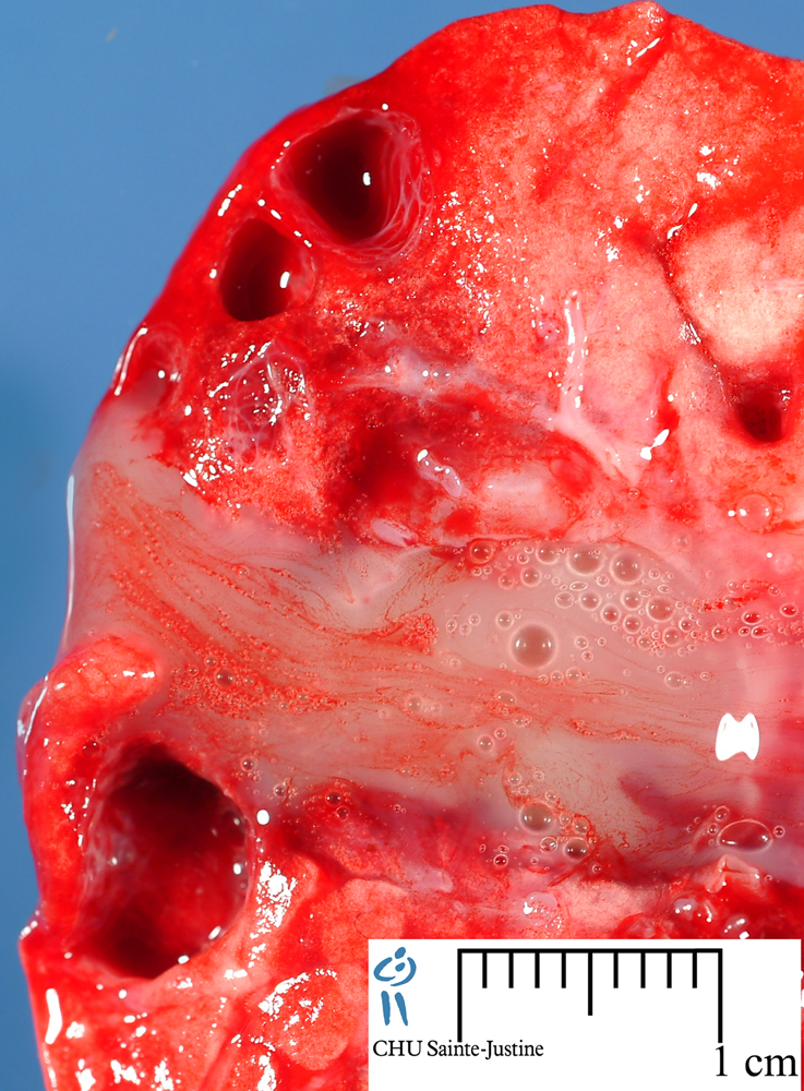
[ (||image_reduire{0,60}|inserer_attribut{alt,Congenital cystic adenomatoid malformation type IV. Macroscopy.}) ] [ (||image_reduire{0,60}|inserer_attribut{alt,CCAM type IV. Macroscopy}) ] [ (||image_reduire{0,60}|inserer_attribut{alt,CCAM type II}) ] [ (||image_reduire{0,60}|inserer_attribut{alt,CPAM type 1}) ] [ (||image_reduire{0,60}|inserer_attribut{alt,CPAM type 1}) ] [ (||image_reduire{0,60}|inserer_attribut{alt,CPAM type 1}) ] [ (||image_reduire{0,60}|inserer_attribut{alt,CPAM type 2 (CCAM type 2)}) ] [ (||image_reduire{0,60}|inserer_attribut{alt,CPAM type 2 (CCAM type 2)}) ] [ (||image_reduire{0,60}|inserer_attribut{alt,CPAM type 2 (CCAM type 2)}) ] [ (||image_reduire{0,60}|inserer_attribut{alt,CPAM type 2 (CCAM type 2)}) ] [ (||image_reduire{0,60}|inserer_attribut{alt,CPAM type 1}) ]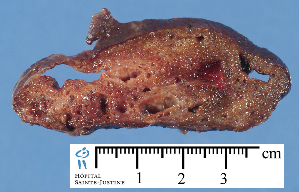
Ent. 1897, Nom. 2002
Digital cases
![]() Case 104 : CPAM type 2
Case 104 : CPAM type 2
Epidemiology
![]() 25% of all congenital lung lesions
25% of all congenital lung lesions
![]() 4-26% of cases can be associated with other congenital abnormalities.
4-26% of cases can be associated with other congenital abnormalities.
![]() estimated incidence: 1 case per 25,000-35,000 pregnancies.
estimated incidence: 1 case per 25,000-35,000 pregnancies.
Subtypes
![]() CPAM type O - CCAM type O (acinar dysplasia)
CPAM type O - CCAM type O (acinar dysplasia)
![]() CPAM type 1 - CCAM type 1 (bronchial/bronchiolar)
CPAM type 1 - CCAM type 1 (bronchial/bronchiolar)
![]() CPAM type 2 - CCAM type 2 (bronchiolar)
CPAM type 2 - CCAM type 2 (bronchiolar)
![]() CPAM type 3 - CCAM type 3 (bronchiolar/alveolar duct)
CPAM type 3 - CCAM type 3 (bronchiolar/alveolar duct)
![]() CPAM type 4 - CCAM type 4 (peripheral)
CPAM type 4 - CCAM type 4 (peripheral)
![]() CPAM type 5 - CCAM type 5
CPAM type 5 - CCAM type 5
CCAM type 0 (congenital acinar dysplasia, congenital acinar aplasia) (1-3%) (neonates, other malformations, poor prognosis)
![]() solid appearance
solid appearance
![]() small and firm lungs
small and firm lungs
![]() bronchial-type airways with cartilage, smooth muscle and glands separated by abundant mesenchymal tissue
bronchial-type airways with cartilage, smooth muscle and glands separated by abundant mesenchymal tissue
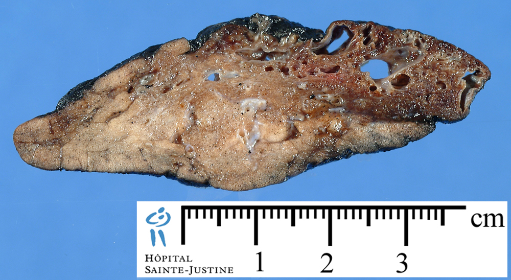
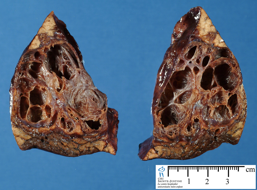
CCAM type 1 (60-70%) (bronchial) (neonates and infants, resectable, good prognosis, possible carcinomatous change)
![]() 1 or more large cysts measuring 2-10 cm in diameter. Larger cysts are often accompanied by smaller cysts, and their walls contain muscle, elastic, or fibrous tissue.
1 or more large cysts measuring 2-10 cm in diameter. Larger cysts are often accompanied by smaller cysts, and their walls contain muscle, elastic, or fibrous tissue.
![]() cartilaginous plates (12%) (#12883247#)
cartilaginous plates (12%) (#12883247#)
![]() Cysts are frequently lined by pseudostratified columnar epithelial cells often interspersed with rows of mucous cells
Cysts are frequently lined by pseudostratified columnar epithelial cells often interspersed with rows of mucous cells
![]() focal mucous cell hyperplasia (12 to 25% of type 1 CCAM)
focal mucous cell hyperplasia (12 to 25% of type 1 CCAM)
![]() microscopic foci of bronchioloalveolar carcinoma (1 to 31% of type 1 CCAM) (#12883247#)
microscopic foci of bronchioloalveolar carcinoma (1 to 31% of type 1 CCAM) (#12883247#)
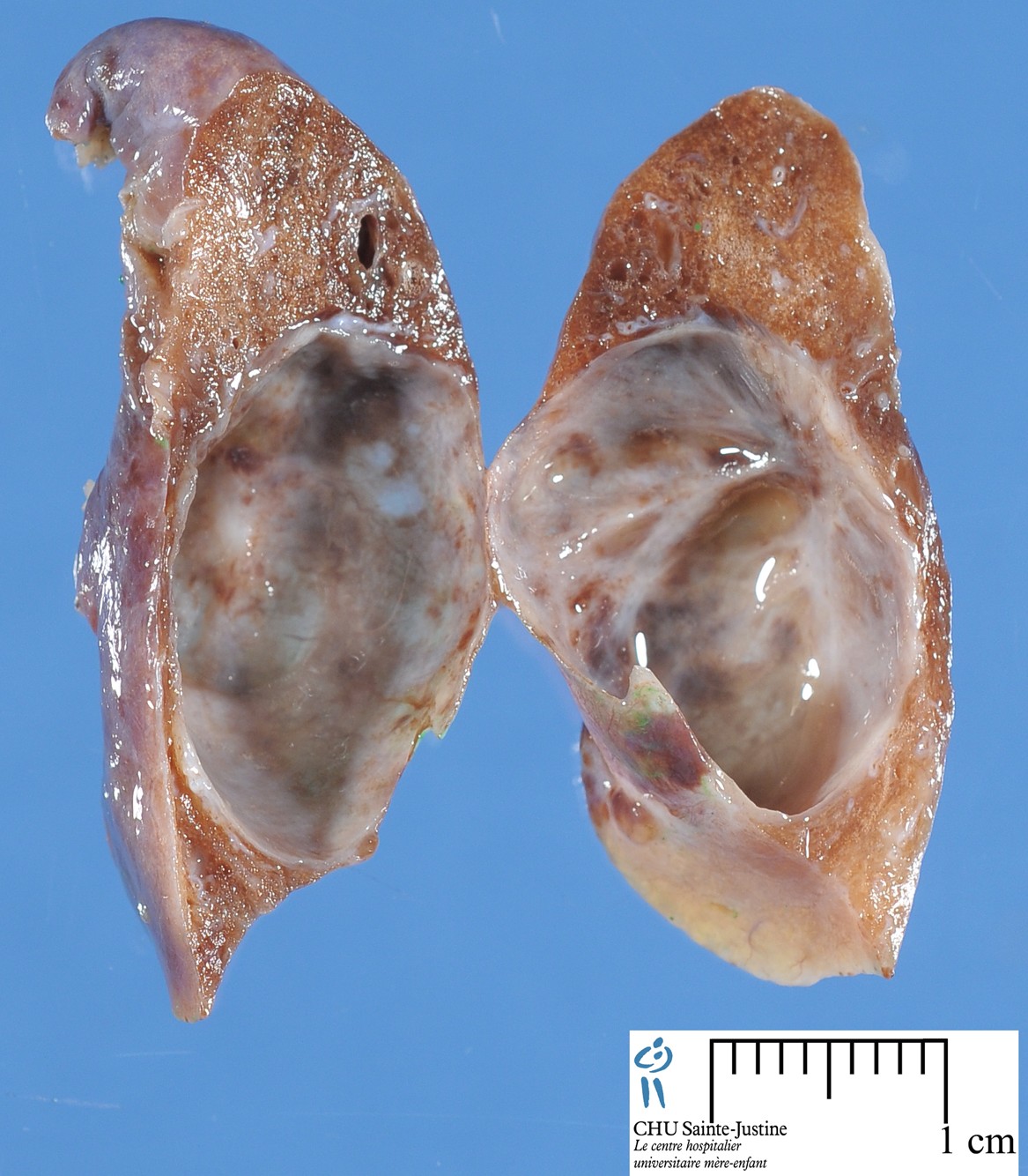
CCAM type 2 (10-15%) (bronchial/bronchiolar) (neonates, other malformation, poor prognosis)
![]() sponge-like appearance
sponge-like appearance
![]() multiple small cysts (0.5 to 2 cm)
multiple small cysts (0.5 to 2 cm)
![]() small relatively uniform cysts resembling bronchioles separated by normal alveoli
small relatively uniform cysts resembling bronchioles separated by normal alveoli
![]() cysts are lined by cuboid-to-columnar epithelium and have a thin fibromuscular wall.
cysts are lined by cuboid-to-columnar epithelium and have a thin fibromuscular wall.
![]() solid pale tumor-like tissue
solid pale tumor-like tissue
![]() striated muscle in 5%
striated muscle in 5%
CCAM type 3 (5%) (bronchiolar) (neonates, poor prognosis)
![]() solid appearance
solid appearance
![]() excess of bronchiolar structure separated by small air spaces, with cuboidal lining, resembling late fetal lung
excess of bronchiolar structure separated by small air spaces, with cuboidal lining, resembling late fetal lung
![]() grossly a solid mass without obvious cyst formation
grossly a solid mass without obvious cyst formation
![]() microscopic adenomatoid cysts
microscopic adenomatoid cysts
CCAM type 4 (28%) (peripheral) (neonates and infants, good prognosis)
![]() large cysts (up to 10 cm)
large cysts (up to 10 cm)
![]() cysts lined by a flattened epithelium (type 1 and 2 pneumocytes) resting on loose mesenchymal tissue
cysts lined by a flattened epithelium (type 1 and 2 pneumocytes) resting on loose mesenchymal tissue
![]() focal stromal hypercellularity (50%) (#12883247#)
focal stromal hypercellularity (50%) (#12883247#)
![]() focal immature cartilage (#12883247#)
focal immature cartilage (#12883247#)
![]() associated pleuropulmonary blastoma (bilateral type 4 CCAM with stromal cellularity) (14%) (#12883247#)
associated pleuropulmonary blastoma (bilateral type 4 CCAM with stromal cellularity) (14%) (#12883247#)
Associations
![]() pulmonary malformations
pulmonary malformations
- extralobar sequestration (CCAM type 2 and CCAM type 3) with systemic arterial supply
- controlateral extralobar pulmonary sequestration (#15300558#)
- bronchial atresia (#7298053#)
- polyalveolar lobe
![]() epithelial hyperplasias
epithelial hyperplasias
- atypical goblet cell hyperplasia
- nonmucinous atypical adenomatous hyperplasia
- focal mucous cell hyperplasia (25% of type 1 CCAM)
![]() renal malformations
renal malformations
- cystic renal disease
- ipsilateral multicystic renal dysplasia (#15630540#)
- bilateral renal agenesis
- contralateral renal agenesis (#15630540#)
![]() ovarian germ cell hypoplasia (#15630540#)
ovarian germ cell hypoplasia (#15630540#)
![]() malignant tumors
malignant tumors
- pleuropulmonary blastoma (bilateral type 4 CCAM with stromal cellularity) (14%) (#12883247#)
- rhabdomyosarcoma (#16007610#, #11431779#, #9314270#)
- malignant mesenchyma
- bronchioloalveolar carcinoma (31% of type 1 CCAM) (#12883247#, #9475333#)
- atypical goblet cell hyperplasia ((#15138930#)
![]() chromosomal anomalies
chromosomal anomalies
- trisomy 13 (#12673637#)
- trisomy 18 (#11173951#)
- chromosme 18 rearrangement (partial deletion of 18p and partial duplication of 18q) (#11173951#)
![]() miscellaneous
miscellaneous
- aneurysm of the vein of Galen (#15022074#)
- sirenomelia
- intestinal atresia
- skeletal malformations
- cleft palate
- hydranencephaly
- hydrocephalus
- diaphragmatic hernia
Differential diagnosis
![]() stromal cellularity in a type 4 CCAM should raise the possibility of blastomatous transformation.
stromal cellularity in a type 4 CCAM should raise the possibility of blastomatous transformation.
See also
![]() congenital pulmonary cysts
congenital pulmonary cysts
![]() pulmonary malformative cysts
pulmonary malformative cysts
![]() pulmonary cystic malformations
pulmonary cystic malformations
![]() pulmonary cyst lesions
pulmonary cyst lesions
References
![]() Riedlinger WF, Vargas SO, Jennings RW, Estroff JA, Barnewolt CE, Lillehei CW, Wilson JM, Colin AA, Reid LM, Kozakewich HP. Bronchial atresia is common to extralobar sequestration, intralobar sequestration, congenital cystic adenomatoid malformation, and lobar emphysema. Pediatr Dev Pathol. 2006 Sep-Oct;9(5):361-73. PMID: #16953677#
Riedlinger WF, Vargas SO, Jennings RW, Estroff JA, Barnewolt CE, Lillehei CW, Wilson JM, Colin AA, Reid LM, Kozakewich HP. Bronchial atresia is common to extralobar sequestration, intralobar sequestration, congenital cystic adenomatoid malformation, and lobar emphysema. Pediatr Dev Pathol. 2006 Sep-Oct;9(5):361-73. PMID: #16953677#
![]() Vargas SO, Korpershoek E, Kozakewich HP, de Krijger RR, Fletcher JA, Perez-Atayde AR. Cytogenetic and p53 profiles in congenital cystic adenomatoid malformation: insights into its relationship with pleuropulmonary blastoma. Pediatr Dev Pathol. 2006 May-Jun;9(3):190-5. PMID: #16944975#
Vargas SO, Korpershoek E, Kozakewich HP, de Krijger RR, Fletcher JA, Perez-Atayde AR. Cytogenetic and p53 profiles in congenital cystic adenomatoid malformation: insights into its relationship with pleuropulmonary blastoma. Pediatr Dev Pathol. 2006 May-Jun;9(3):190-5. PMID: #16944975#
![]() Wilson RD, Hedrick HL, Liechty KW, Flake AW, Johnson MP, Bebbington M, Adzick NS. Cystic adenomatoid malformation of the lung: Review of genetics, prenatal diagnosis, and in utero treatment. Am J Med Genet A. 2006 Jan 15;140(2):151-5. PMID: #16353256#
Wilson RD, Hedrick HL, Liechty KW, Flake AW, Johnson MP, Bebbington M, Adzick NS. Cystic adenomatoid malformation of the lung: Review of genetics, prenatal diagnosis, and in utero treatment. Am J Med Genet A. 2006 Jan 15;140(2):151-5. PMID: #16353256#
![]() MacSweeney F, Papagiannopoulos K, Goldstraw P, Sheppard MN, Corrin B, Nicholson AG. An assessment of the expanded classification of congenital cystic adenomatoid malformations and their relationship to malignant transformation. Am J Surg Pathol. 2003 Aug;27(8):1139-46. PMID: #12883247#
MacSweeney F, Papagiannopoulos K, Goldstraw P, Sheppard MN, Corrin B, Nicholson AG. An assessment of the expanded classification of congenital cystic adenomatoid malformations and their relationship to malignant transformation. Am J Surg Pathol. 2003 Aug;27(8):1139-46. PMID: #12883247#
![]() Stocker JT, Madewell JE, Drake RM. Congenital cystic adenomatoid malformation of the lung. Classification and morphologic spectrum. Hum Pathol. 1977 Mar;8(2):155-71. PMID: #856714#
Stocker JT, Madewell JE, Drake RM. Congenital cystic adenomatoid malformation of the lung. Classification and morphologic spectrum. Hum Pathol. 1977 Mar;8(2):155-71. PMID: #856714#
![]() Cha I, Adzick NS, Harrison MR, Finkbeiner WE. Fetal congenital cystic adenomatoid malformations of the lung: a clinicopathologic study of eleven cases. Am J Surg Pathol. 1997 May;21(5):537-44. PMID: #9158677#
Cha I, Adzick NS, Harrison MR, Finkbeiner WE. Fetal congenital cystic adenomatoid malformations of the lung: a clinicopathologic study of eleven cases. Am J Surg Pathol. 1997 May;21(5):537-44. PMID: #9158677#
![]() Cangiarella J, Greco MA, Askin F, et al. Congenital cystic adenomatoid malformation of the lung: insights into the pathogenesis utilizing quantitative analysis of vascular marker CD34 (QBEND-10) and cell proliferation marker MIB-1. Mod Pathol 1995; 8:913-8.
Cangiarella J, Greco MA, Askin F, et al. Congenital cystic adenomatoid malformation of the lung: insights into the pathogenesis utilizing quantitative analysis of vascular marker CD34 (QBEND-10) and cell proliferation marker MIB-1. Mod Pathol 1995; 8:913-8.