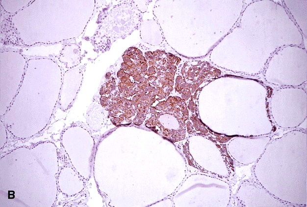
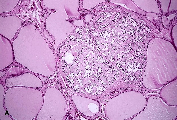
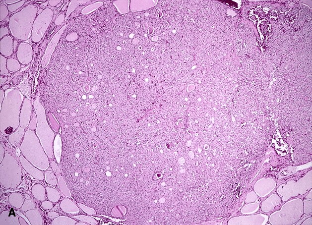
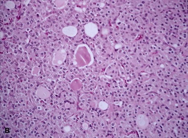
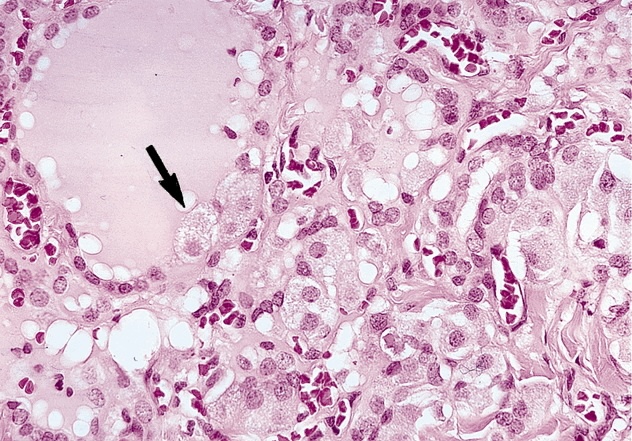
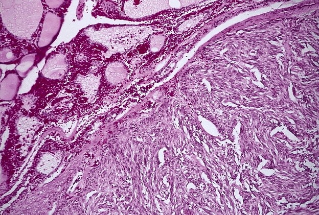
microscopic medullary thyroid carcinoma
Image Gallery
[ (||image_reduire{0,60}|inserer_attribut{alt,Microscopic medullary thyroid carcinoma (PMID:9630179)}) ] [ (||image_reduire{0,60}|inserer_attribut{alt,Microscopic medullary thyroid carcinoma (PMID:10843288)}) ] [ (||image_reduire{0,60}|inserer_attribut{alt,Microscopic medullary thyroid carcinoma (PMID:10843288)}) ] [ (||image_reduire{0,60}|inserer_attribut{alt,Microscopic medullary thyroid carcinoma (PMID:10843288)}) ] [ (||image_reduire{0,60}|inserer_attribut{alt,Microscopic medullary thyroid carcinoma (PMID:10843288)}) ] [ (||image_reduire{0,60}|inserer_attribut{alt,Amyloid microscopic medullary thyroid carcinoma (PMID:10843288)}) ] [ (||image_reduire{0,60}|inserer_attribut{alt,C-cell hyperplasia with amyloid microscopic medullary thyroid carcinoma (...)}) ] [ (||image_reduire{0,60}|inserer_attribut{alt,Microscopic medullary thyroid carcinoma with follicular pattern (...)}) ] [ (||image_reduire{0,60}|inserer_attribut{alt,Microscopic medullary thyroid carcinoma with follicular pattern (...)}) ] [ (||image_reduire{0,60}|inserer_attribut{alt,C-cell hyperplasia with microscopic medullary thyroid carcinoma (...)}) ] [ (||image_reduire{0,60}|inserer_attribut{alt,Spindle cell-microscopic medullary thyroid carcinoma (PMID:10843288)}) ]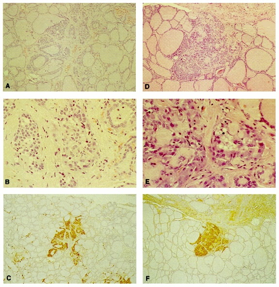
Microcarcinomas of the thyroid are defined as tumors measuring less than 1.0 cm in greatest dimension.
Medullary thyroid microcarcinomas have been described previously as incidental surgical or autopsy findings, with an autopsy incidence of 0.15%.
Only rarely have symptomatic sporadic or genetically determined medullary thyroid microcarcinomas been reported.
Differential diagnosis
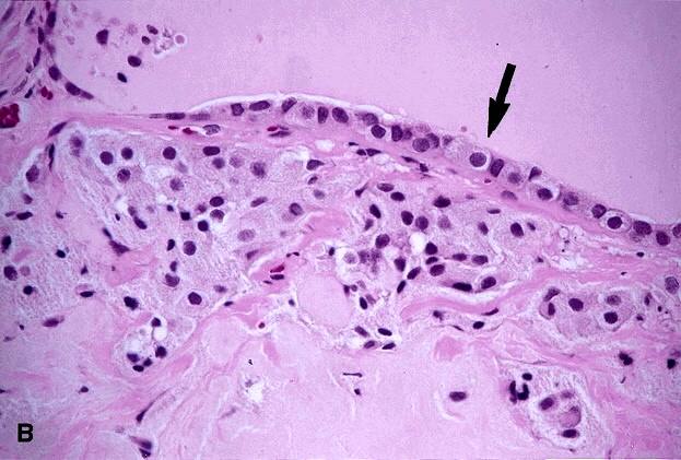
At a light microscopic level, the separation of C-cell hyperplasia and microscopic medullary carcinoma of the thyroid (MCT) is difficult, and it ultimately rests on the finding of C cells outside of the thyroid follicular basement membranes (FBMs).
Types
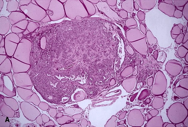
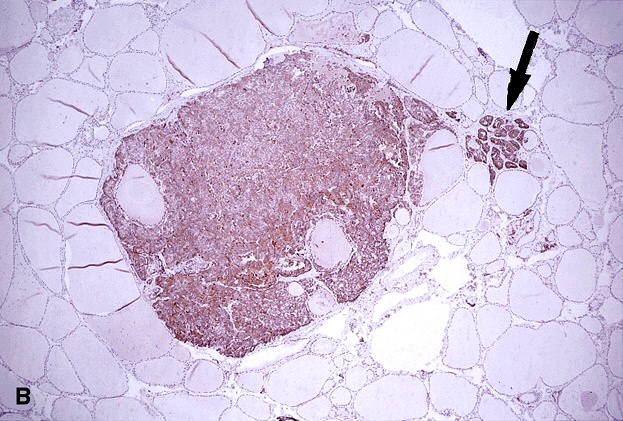
![]() unifocal microscopic MCT
unifocal microscopic MCT
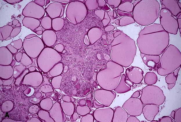
![]() multifocal microscopic MCT
multifocal microscopic MCT
Collagen type IV (CIV) immunostaining (#8522302#)
Three patterns of C-cell proliferation can be recognized in the thyroid follicular basement membranes by collagen type IV (CIV). (#8522302#)
![]() The first is characterized by complete investment of C-cells by a continuous rim of collagen type IV (CIV), corresponding to the thyroid follicular basement membranes (FBM) and confirming an intrafollicular localization; hence, the diagnosis of C-cell hyperplasia (CCH) is made in such cases.
The first is characterized by complete investment of C-cells by a continuous rim of collagen type IV (CIV), corresponding to the thyroid follicular basement membranes (FBM) and confirming an intrafollicular localization; hence, the diagnosis of C-cell hyperplasia (CCH) is made in such cases.
![]() The second pattern is distinctive and is typified by defects in the collagen type IV (CIV) layer; constituent C-cells assumed an extrafollicular location. These images yielded a diagnosis of microscopic-MCT.
The second pattern is distinctive and is typified by defects in the collagen type IV (CIV) layer; constituent C-cells assumed an extrafollicular location. These images yielded a diagnosis of microscopic-MCT.
![]() The third findings are also accompanied by focal reduplication of basement membrane that is apparently tumor derived, producing a micronodular or microlobular configuration. The third pattern represents a combination of the first two, with C-cell nodules that were bounded by CIV and clearly situated in an intrafollicular location; however, focal reduplication of basement membranes was also evident in these cases. The biological significance of the third pattern of collagen type IV (CIV) staining is uncertain, but it may reflect the presence of a preinvasive proliferation of C-cells, that is distinct from "usual" CCH in MEN2a.
The third findings are also accompanied by focal reduplication of basement membrane that is apparently tumor derived, producing a micronodular or microlobular configuration. The third pattern represents a combination of the first two, with C-cell nodules that were bounded by CIV and clearly situated in an intrafollicular location; however, focal reduplication of basement membranes was also evident in these cases. The biological significance of the third pattern of collagen type IV (CIV) staining is uncertain, but it may reflect the presence of a preinvasive proliferation of C-cells, that is distinct from "usual" CCH in MEN2a.
See also
![]() multifocal C-cell hyperplasia
multifocal C-cell hyperplasia
- diffuse C-cell hyperplasia
- nodular C-cell hyperplasia
![]() medullary thyroid carcinoma
medullary thyroid carcinoma
References
![]() Sporadic versus familial medullary thyroid microcarcinoma: a histopathologic study of 50 consecutive patients. Kaserer K, Scheuba C, Neuhold N, Weinhäusel A, Haas OA, Vierhapper H, Niederle B. Am J Surg Pathol. 2001 Oct;25(10):1245-51. PMID: #11688458#
Sporadic versus familial medullary thyroid microcarcinoma: a histopathologic study of 50 consecutive patients. Kaserer K, Scheuba C, Neuhold N, Weinhäusel A, Haas OA, Vierhapper H, Niederle B. Am J Surg Pathol. 2001 Oct;25(10):1245-51. PMID: #11688458#
![]() Inherited medullary microcarcinoma of the thyroid: a study of 11 cases. Krueger JE, Maitra A, Albores-Saavedra J. Am J Surg Pathol. 2000 Jun;24(6):853-8. PMID: #10843288#
Inherited medullary microcarcinoma of the thyroid: a study of 11 cases. Krueger JE, Maitra A, Albores-Saavedra J. Am J Surg Pathol. 2000 Jun;24(6):853-8. PMID: #10843288#
![]() McDermott MB, Swanson PE, Wick MR. Immunostains for collagen type IV discriminate between C-cell hyperplasia and microscopic medullary carcinoma in multiple endocrine neoplasia, type 2a. Hum Pathol 1995;26:1308-312. PMID: #8522302#
McDermott MB, Swanson PE, Wick MR. Immunostains for collagen type IV discriminate between C-cell hyperplasia and microscopic medullary carcinoma in multiple endocrine neoplasia, type 2a. Hum Pathol 1995;26:1308-312. PMID: #8522302#