fibrous cortical defect
Image Gallery
[ (||image_reduire{0,60}|inserer_attribut{alt,Fibrous cortical defect (eMedicine)}) ] [ (||image_reduire{0,60}|inserer_attribut{alt,Fibrous cortical defect (eMedicine)}) ] [ (||image_reduire{0,60}|inserer_attribut{alt,Fibrous cortical defect (eMedicine)}) ] [ (||image_reduire{0,60}|inserer_attribut{alt,Fibrous cortical defect (eMedicine)}) ] [ (||image_reduire{0,60}|inserer_attribut{alt,Multiple fibrous cortical defects}) ]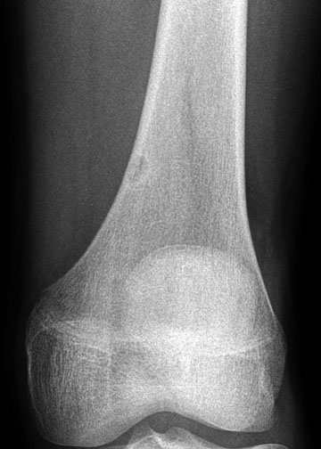
The fibrous cortical defects (FCDs) have been mislabeled or described as cortical avulsive irregularities, subperiosteal or periosteal desmoid, a variant of periostitis ossificans, and cortical desmoid.
In the literature, fibrous cortical defect (FCD) has a long list of pseudonyms, including metaphyseal fibrous defect, metaphyseal supracondylar cortical defect, and developmental defect.
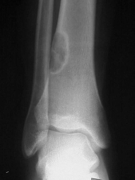
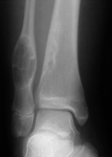
The non-ossifying fibroma (NOF or fibroxanthoma) and fibrous cortical defects (FCDs) are nonaggressive fibrous lesions of bone that are distinguished from one another historically by their size and natural history.
Both are considered to be developmental defects and to be nonaggressive. They were first described by Lichtenstein and Jaffe in 1942 and they typically occurred within the metaphysis of growing long tubular bones in children, most commonly about the knee. Controversy continues regarding the correct terminology and etiology of these lesions.
The distinction between fibrous cortical defect (FCD) and non-ossifying fibroma (NOF) is based on size and natural history.
Fibrous cortical defects (FCDs) are asymptomatic, small (<3 cm), eccentrically located, metaphyseal cortical defects; most of these spontaneously disappear. However, some evolve and enlarge into non-ossifying fibromas (NOFs).
Conversely, non-ossifying fibromas are larger (>3 cm), eccentric, intramedullary lesions that abut the cortex; they have a typical, superficial, scalloping pattern in the adjacent cortex.
While these lesions also can heal spontaneously (with reactive bone filling in the central lucent fibrous tissue component), they can also persist, with interval growth that continues into adulthood.
Typically, non-ossifying fibromas (NOFs) are asymptomatic. However, the larger lesions may become symptomatic, with a risk of pathologic fracture. Steiner suggested that these 2 lesions are secondary to cellular proliferation due to aberrations in local development.
The group non-ossifying fibroma (NOF), fibrous cortical defect (FCD), fibrous metaphyseal defect, benign fibrous histiocytoma
The terms fibroxanthoma, nonossifying fibroma (NOF), fibrous cortical defect (FCD), and, less commonly, benign fibrous histiocytoma have all been used interchangeably in the radiology literature.
The non-ossifying fibroma (NOF) and the fibrous cortical defect (FCD), however, are considered to be 2 distinct lesions with respect to size and natural history.
The term "Fibroxanthoma" has been the preferred for the non-ossifying fibroma (NOF) lesion because it more accurately reflects the underlying pathologic findings.
Differential diagnosis
The cortical desmoid—or cortical avulsive injury, as it is currently known—is believed to be separate from the cortical defect, and it has been related to repetitive stress at the attachment of the extensor tendon fibers along the linea aspera of the distal femur.
Treatment and prognosis
No specific treatment or intervention is required for FCDs, and they are usually left alone. They may persist into adulthood without complications and eventually become non-ossifying fibromas (NOFs or fibroxanthomas). However, if they are removed at biopsy, they do not recur.
With respect to fibroxanthomas, small asymptomatic lesions do not require biopsy or treatment. With larger lesions, careful radiographic observation and decreased vigorous activity of the patient are recommended.
Curettage and bone graft procedures are performed to prevent a pathologic fracture if the lesion becomes larger than 33 mm in diameter or involves more than 50% of the transverse diameter of a critical weight-bearing bone.
Links
See also
![]() non-ossifying fibroma
non-ossifying fibroma
![]() fibrous metaphyseal defect
fibrous metaphyseal defect
![]() benign fibrous histiocytoma of bone
benign fibrous histiocytoma of bone