Home > G. Tumoral pathology > perivascular epithelioid cell tumor
perivascular epithelioid cell tumor
Monday 15 March 2004
PEComa; PEComas; clear cell ’sugar’ tumor, clear cell myomelanocytic tumor. Angiomyolipoma, lymphangioleiomyomatosis, clear cell sugar tumor of the lung, clear cell myomelanocytic tumor of ligamentum teres/falciform ligament, and abdominopelvic sarcoma of perivascular epithelioid cells.
Ent, 1992; Nom, 1992
| WKP | PO |
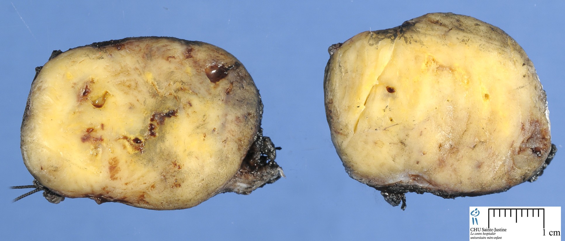
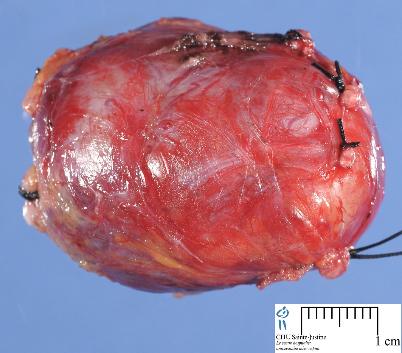
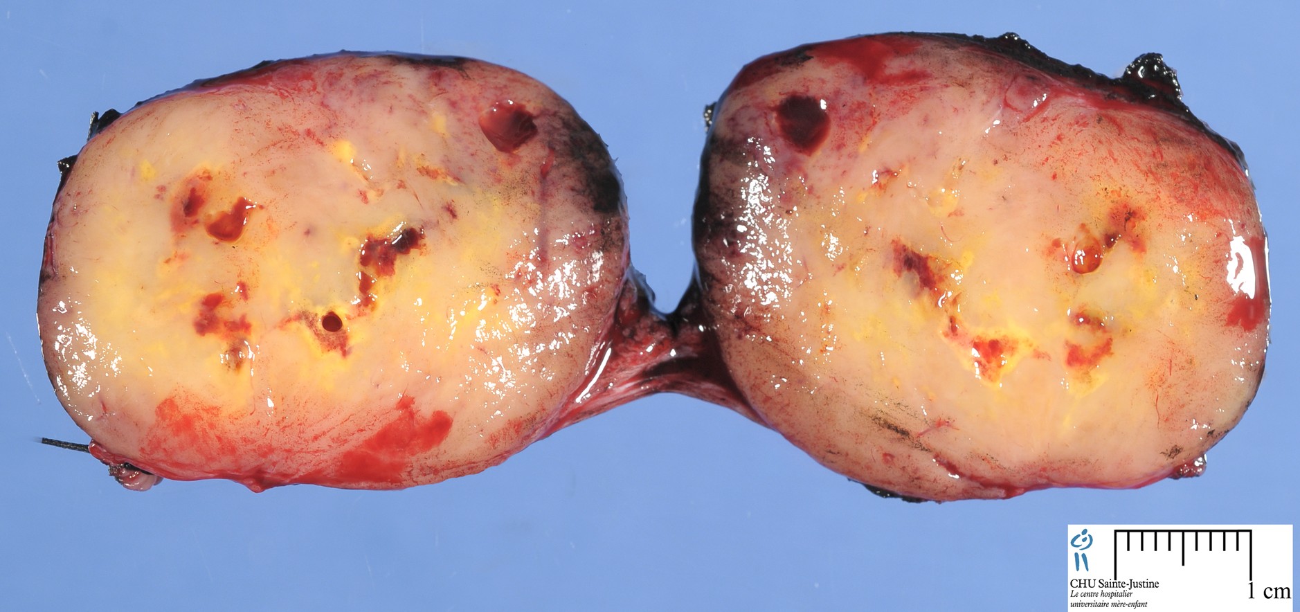
Definition: Perivascular epithelioid cell neoplasms (PEComa) are a family of rare mesenchymal tumors with hybrid myo-melanocytic differentiation. Although most PEComas harbor loss-of-function TSC1/TSC2 mutations, a small subset were reported to carry TFE3 gene rearrangements.
Perivascular epithelioid cell tumour, also known as PEComa or PEC tumour, is a family of mesenchymal tumours consisting of perivascular epithelioid cells (PECs). These are rare tumours that can occur in any part of the human body.
See also
Images
PEComa
Malignant PEComa
- https://twitter.com/JMGardnerMD/status/678367493937737729
- https://twitter.com/JMGardnerMD/status/678030539601797120
The family of tumors derived from mesenchymal perivascular epithelioid cells (so-called PEComas) includes angiomyolipoma, lymphangioleiomyomatosis, clear cell sugar tumor of the lung, clear cell myomelanocytic tumor of ligamentum teres/falciform ligament, and abdominopelvic sarcoma of perivascular epithelioid cells. These tumors were characterized by coexpression of melanocytic (HMB-45) and muscle markers.
The precise nature of PEComa is still poorly understood. This is a morphological spectrum of lesions composed of perivascular epithelioid or more spindled cells, showing variably clear or granular eosinophilic cytoplasm and a variable degree of pleomorphism.
The tumour cells are very often oriented closely around prominent thin-walled blood vessels. Distinctively, these lesions appear to coexpress markers of both myoid and melanocytic differentiation.
Angiomyolipoma, clear cell (sugar) tumour and lymphangiomyomatosis belong in this category but retain their existing names if morphologically and topographically typical.
Instead, the term PEComa is generally used for lesions arising most often in soft tissue or in visceral locations outside the kidney, liver or lung.
While data are limited thus far, many (perhaps most) of these tumours pursue a benign course but a subset behave in a malignant fashion, often in the context of more pleomorphic epithelioid cytomorphology.
Members
angiomyolipoma
lymphangioleiomyomatosis
clear cell sugar tumor of the lung
clear cell myomelanocytic tumor of ligamentum teres/falciform ligament (10976698)
abdominopelvic sarcoma of perivascular epithelioid cells
Localization
digestive PEComas (15614743)
common bile duct (15252321)
uterus (16327428, 15213607, 15043315, 13678746)
uterine cervix (15494070)
prostate (12562263)
tigh (11756764)
falciform ligament (10976698)
skin : cutaneous PEComa (15842631, 18277881)
skull base (15316324)
soft tissue (15577688)
- orbit (15803216)
- broad ligament (15361222)
- breast (11979098)
Variants
malignant perivascular epithelioid cell tumor
- https://twitter.com/JMGardnerMD/status/678367493937737729
- Malignant PEComa mimics alveolar soft part sarcoma (ASPS)
Immunochemistry
HMB-45+
progesterone receptor+
MyoD1+ (12778001)
CK-
EMA-
melan-A-
S100-
Molecular biology
loss-of-function TSC1/TSC2 mutations
SFPQ/PSF-TFE3 Gene Fusion (19606011): 23%
- TFE3-associated PEComas
RAD51B gene rearrangements were identified in uterine PEComas (8%)
HTR4-ST3GAL1 fusion
RASSF1-PDZRN3 fusion
In a study (25651471), combined RNA sequencing and fluorescence in situ hybridization analysis identified 9 (23%) TFE3 gene-rearranged tumors, with 3 cases showing an SFPQ/PSF-TFE3 fusion and 1 case showing a novel DVL2-TFE3 gene fusion. The TFE3-positive lesions showed a distinctive nested/alveolar morphology and were equally distributed between soft tissue and visceral sites.
In addition, novel RAD51B gene rearrangements were identified in 3 (8%) uterine PEComas, which showed a complex fusion pattern and were fused to RRAGB/OPHN1 genes in 2 cases. Other nonrecurrent gene fusions, HTR4-ST3GAL1 and RASSF1-PDZRN3, were identified in 2 cases.
TSC2 mutations were identified in 80% of the TFE3 fusion-negative cases tested. Coexistent TP53 mutations were identified in 63% of the TSC2-mutated PEComas.
These results showed that TFE3-rearranged PEComas lacked coexisting TSC2 mutations, indicating alternative pathways of tumorigenesis. (25651471)
See also
Tumors
- clear cell tumors
- clear cell carcinomas
- clear cell sarcomas
References
Dichotomy of Genetic Abnormalities in PEComas With Therapeutic Implications. Agaram NP, Sung YS, Zhang L, Chen CL, Chen HW, Singer S, Dickson MA, Berger MF, Antonescu CR. Am J Surg Pathol. 2015 Feb 3. PMID: 25651471
Perivascular Epithelioid Cell Tumor With SFPQ/PSF-TFE3 Gene Fusion in a Patient With Advanced Neuroblastoma. Tanaka M, Kato K, Gomi K, Matsumoto M, Kudo H, Shinkai M, Ohama Y, Kigasawa H, Tanaka Y. Am J Surg Pathol. 2009 Jul 13. PMID: 19606011
Iyengar P, Deangelis DD, Greenberg M, Taylor G. Perivascular epithelioid cell tumor of the orbit: a case report and review of the literature. Pediatr Dev Pathol. 2005 Jan-Feb;8(1):98-104. PMID: 15803216
Vang R, Kempson RL. Perivascular epithelioid cell tumor (’PEComa’) of the uterus: a subset of HMB-45-positive epithelioid mesenchymal neoplasms with an uncertain relationship to pure smooth muscle tumors. Am J Surg Pathol. 2002 Jan;26(1):1-13. PMID: 11756764
Tazelaar HD, Batts KP, Srigley JR. Primary extrapulmonary sugar tumor (PEST): a report of four cases. Mod Pathol. 2001 Jun;14(6):615-22. PMID: 11406665
Folpe AL, Goodman ZD, Ishak KG, Paulino AF, Taboada EM, Meehan SA, Weiss SW. Clear cell myomelanocytic tumor of the falciform ligament/ligamentum teres: a novel member of the perivascular epithelioid clear cell family of tumors with a predilection for children and young adults. Am J Surg Pathol. 2000 Sep;24(9):1239-46. PMID: 10976698