Home > G. Tumoral pathology > infantile myofibroma
infantile myofibroma
Tuesday 9 December 2003
Digital case
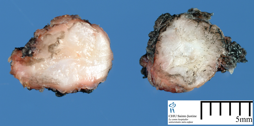
![]() Case 205 (HPC:205) : Infantile myofibroma
Case 205 (HPC:205) : Infantile myofibroma
Epidemiology
![]() sporadic cases
sporadic cases![]() familial cases (8600777)
familial cases (8600777)
- autosomal dominant inheritance (12894106, 1739928, 6742314)
- autosomal-recessive inheritance (11260217)
Localization of tumors
![]() orbital region (11410140)
orbital region (11410140)![]() central nervous system (12720031), spinal canal (9703012)
central nervous system (12720031), spinal canal (9703012)![]() oral region (10792791), gingiva (12472999)
oral region (10792791), gingiva (12472999)![]() bone
bone![]() muscle
muscle![]() viscera (heart, lungs, liver, gastrointestinal tract (2672792), bile ducts and pancreas (3185365), endocrine organs)
viscera (heart, lungs, liver, gastrointestinal tract (2672792), bile ducts and pancreas (3185365), endocrine organs)![]() subcutaneous tissue, soft tissues
subcutaneous tissue, soft tissues ![]() digestive tract
digestive tract
- intestinal infantile myofibromatosis
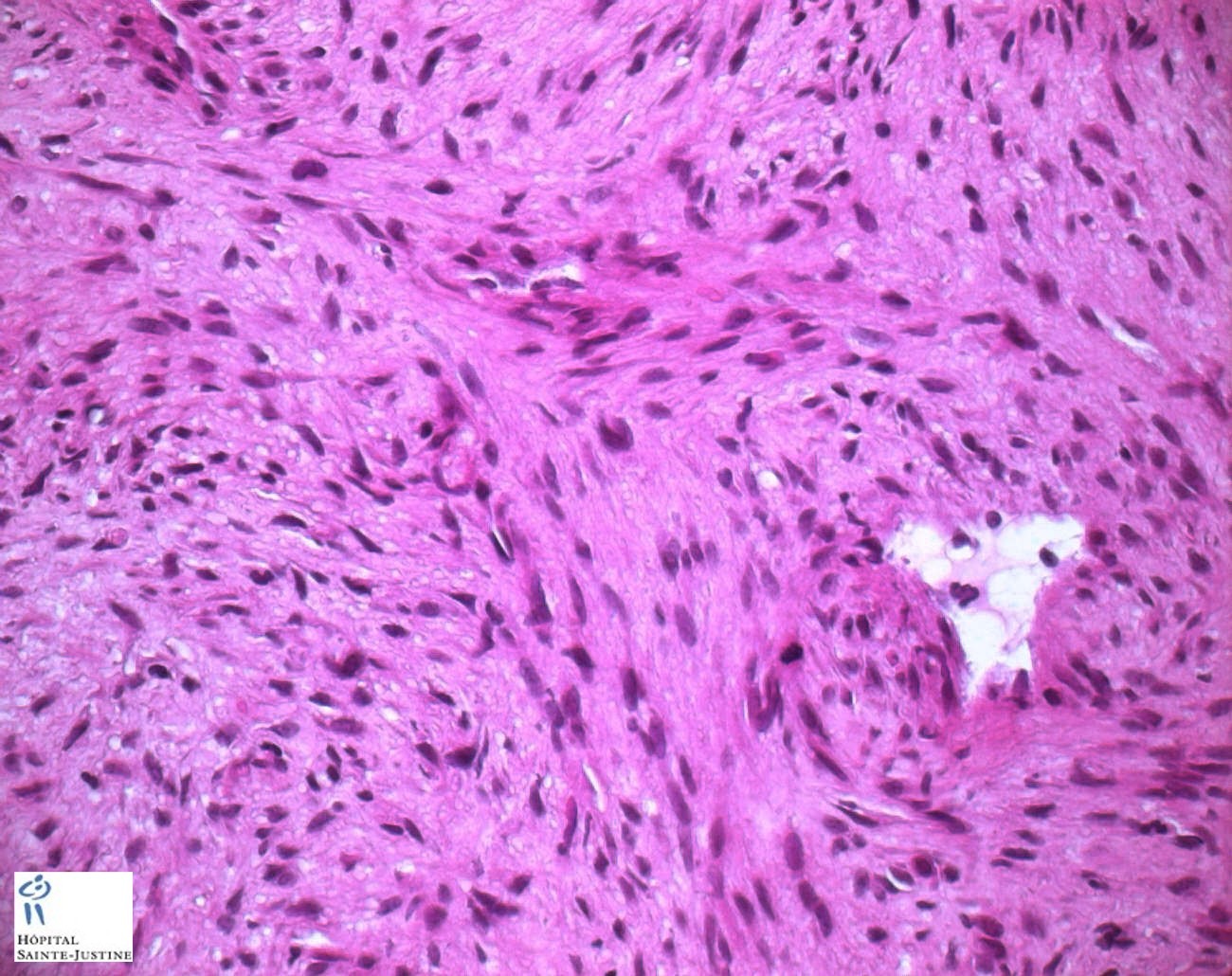
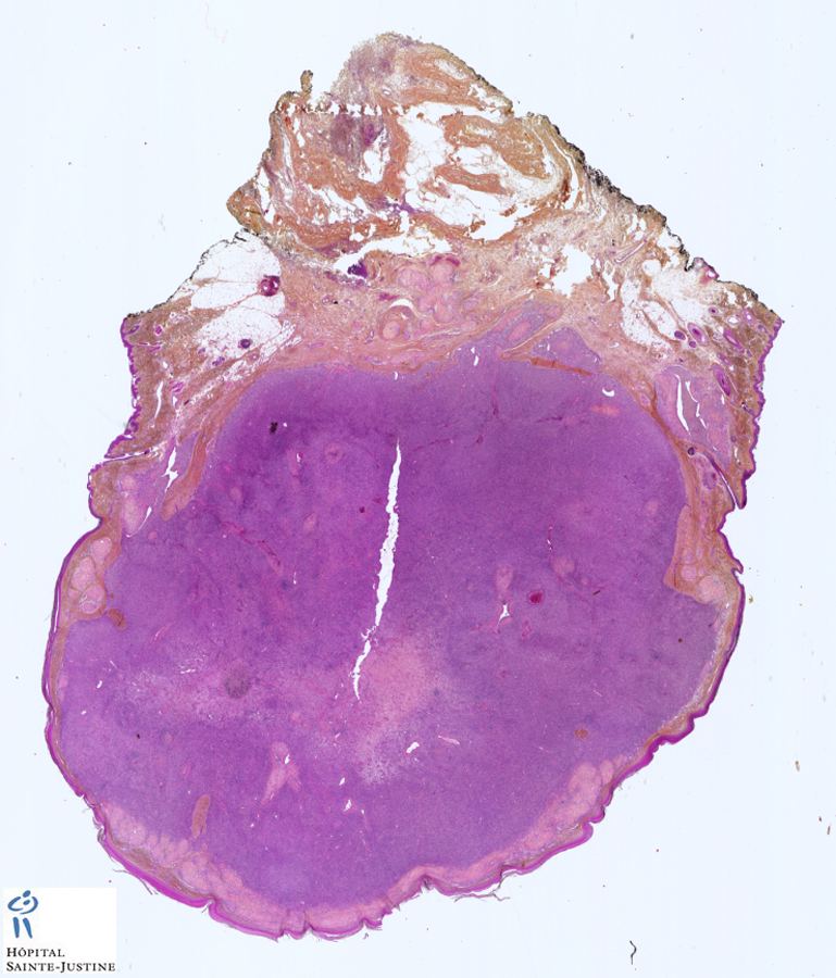
Microscopical synopsis
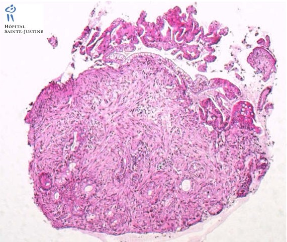
Biphasic pattern![]() fascicles of spindle cells with abundant eosinophilic cytoplasm that resembled smooth muscle
fascicles of spindle cells with abundant eosinophilic cytoplasm that resembled smooth muscle![]() more primitive spindled cells associated with a hemangiopericytoma-like vascular pattern
more primitive spindled cells associated with a hemangiopericytoma-like vascular pattern
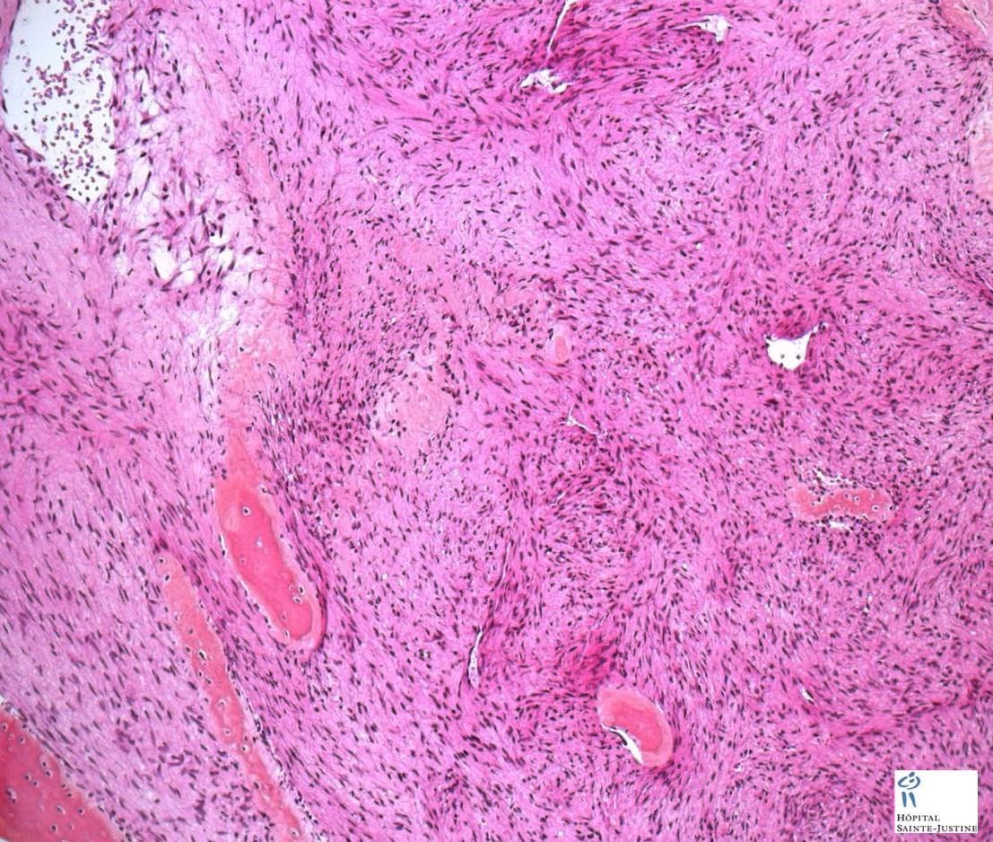
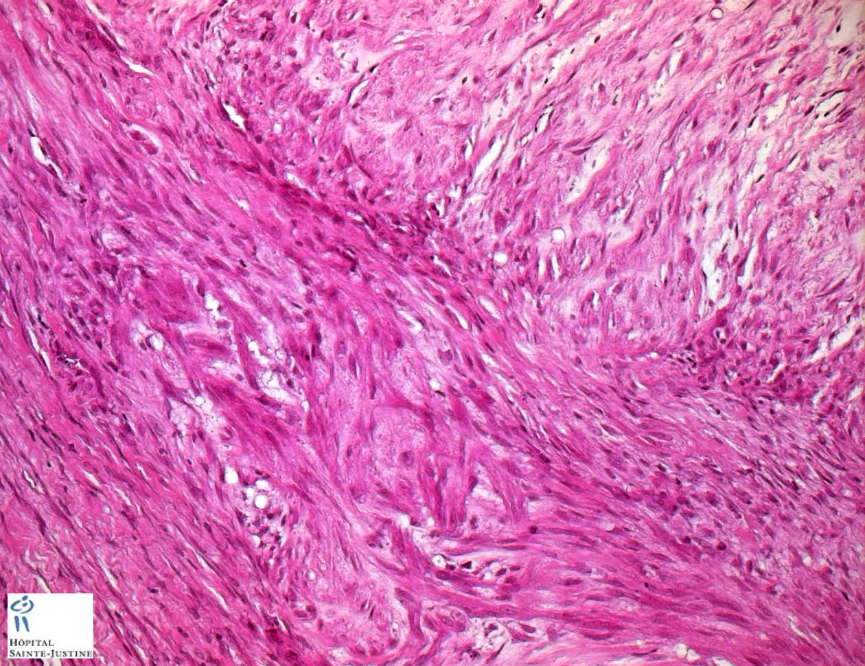
![]() interlacing fascicles of myofibroblasts with abundant eosinophilic cytoplasm
interlacing fascicles of myofibroblasts with abundant eosinophilic cytoplasm ![]() variable necrosis
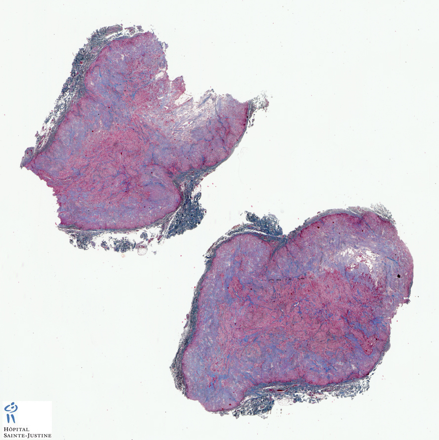
variable necrosis ![]() calcifications in some sites
calcifications in some sites![]() hemangiopericytoma-like features
hemangiopericytoma-like features ![]() angiocentric and perivascular growth of myofibroblasts (+/-) (8597844)
angiocentric and perivascular growth of myofibroblasts (+/-) (8597844)
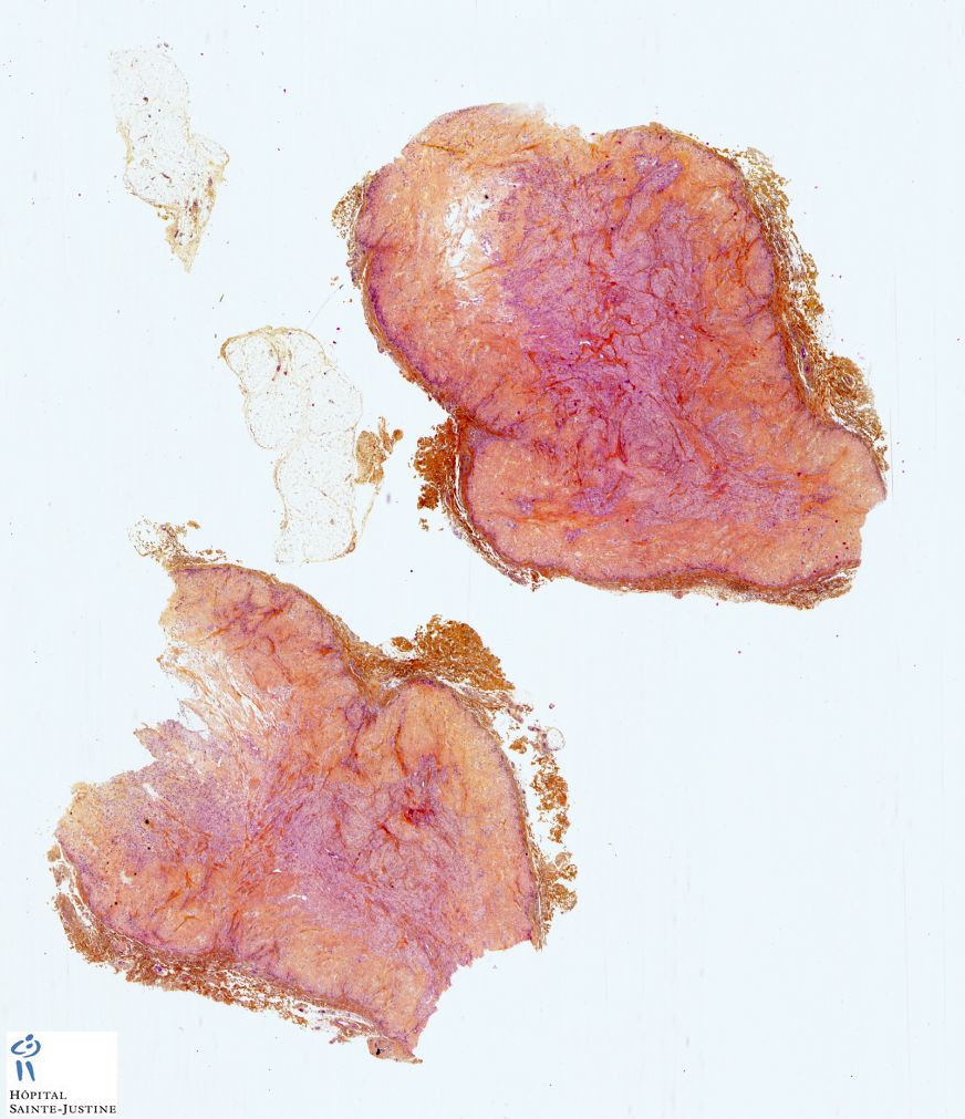
Immunochemistry
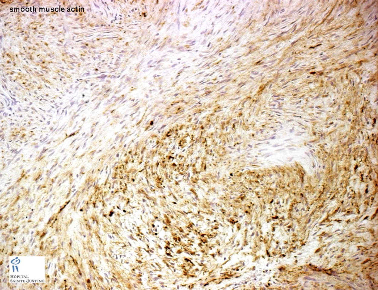
![]() vimentin+
vimentin+![]() smooth muscle actin+
smooth muscle actin+
Subtypes
![]() solitary myofibromatosis
solitary myofibromatosis![]() systemic myofibromatosis, multicentric myofibromatosis
systemic myofibromatosis, multicentric myofibromatosis
- congenital generalized myofibromatosis (CGMF) (8597844)
- aggressive infantile myofibromatosis (3784986)
Two types can be distinguished; the solitary type, defined by the presence of one nodule in the skin, muscle, bone or subcutaneous tissue; and the multicentric type which can be divided into two sub-types.
In the first sub-type the lesions are multicentric but without visceral involvement, while in the second, visceral involvement is present.
![]() adult-type myofibroma
adult-type myofibroma
Associations (sporadic)
![]() neonatal hemochromatosis (11196755)
neonatal hemochromatosis (11196755)![]() Turner syndrome (9821432)
Turner syndrome (9821432)![]() arterial fibromuscular dysplasia (15455480)
arterial fibromuscular dysplasia (15455480)
Cytogenetics
![]() del(6)(q12q15) (10425309)
del(6)(q12q15) (10425309)![]() monosomy 9q (15365831)
monosomy 9q (15365831)![]() trisomy 16q (15365831)
trisomy 16q (15365831)
Prognosis
The prognosis of the disease depends on whether visceral involvement is present. Solitary and multicentric nodules without visceral involvement usually have excellent prognosis with spontaneous regression of lesions within 1 to 2 years of diagnosis.
Visceral lesions are associated with a significant morbidity and mortality, resulting from vital organ obstruction, failure to thrive, or infection. Death in these cases often occurs at birth, or soon after, and is usually due to cardio-pulmonary or gastrointestinal complications. However, multicentic type of infantile myofibromatosis with visceral involvement can spontaneously regress. (11510506)
Differential diagnosis
![]() low-grade myofibroblastic neoplasms
low-grade myofibroblastic neoplasms
![]() fibromatoses
fibromatoses
- composite fibromatoses
![]() congenital fibrosarcoma (infantile fibrosarcoma)
congenital fibrosarcoma (infantile fibrosarcoma)![]() inflammatory myofibroblastic tumor
inflammatory myofibroblastic tumor![]() fibrous fibrohistiocytic tumors
fibrous fibrohistiocytic tumors![]() solitary fibrous tumor
solitary fibrous tumor![]() nodular fasciitis
nodular fasciitis![]() desmoplastic fibroblastoma (collagenous fibroma)
desmoplastic fibroblastoma (collagenous fibroma)![]() smooth muscle tumors
smooth muscle tumors
- leiomyoma
- leiomyosarcoma
![]() neurogenic tumors
neurogenic tumors
- schwannoma and neurofibroma
- low-grade MPNST
![]() juvenile xanthogranuloma
juvenile xanthogranuloma
See also
![]() hemaniopericytomatous pattern
hemaniopericytomatous pattern
References
![]() Granter SR, Badizadegan K, Fletcher CD. Myofibromatosis in adults, glomangiopericytoma, and myopericytoma: a spectrum of tumors showing perivascular myoid differentiation. Am J Surg Pathol. 1998 May;22(5):513-25. PMID: 9591720
Granter SR, Badizadegan K, Fletcher CD. Myofibromatosis in adults, glomangiopericytoma, and myopericytoma: a spectrum of tumors showing perivascular myoid differentiation. Am J Surg Pathol. 1998 May;22(5):513-25. PMID: 9591720
![]() Mentzel T, Calonje E, Nascimento AG, Fletcher CD. Infantile hemangiopericytoma versus infantile myofibromatosis. Study of a series suggesting a continuous spectrum of infantile myofibroblastic lesions. Am J Surg Pathol. 1994 Sep;18(9):922-30. PMID: 8067513
Mentzel T, Calonje E, Nascimento AG, Fletcher CD. Infantile hemangiopericytoma versus infantile myofibromatosis. Study of a series suggesting a continuous spectrum of infantile myofibroblastic lesions. Am J Surg Pathol. 1994 Sep;18(9):922-30. PMID: 8067513
![]() Dictor M, Elner A, Andersson T, Ferno M. Myofibromatosis-like hemangiopericytoma metastasizing as differentiated vascular smooth-muscle and myosarcoma. Myopericytes as a subset of "myofibroblasts". Am J Surg Pathol. 1992 Dec;16(12):1239-47. PMID: 1463097
Dictor M, Elner A, Andersson T, Ferno M. Myofibromatosis-like hemangiopericytoma metastasizing as differentiated vascular smooth-muscle and myosarcoma. Myopericytes as a subset of "myofibroblasts". Am J Surg Pathol. 1992 Dec;16(12):1239-47. PMID: 1463097