Home > D. General pathology > Blood and immunity > Thymus > thymoma type A
thymoma type A
Friday 7 December 2012
Spindle cell thymoma. Thymoma, type A.
Definition: Type A thymoma (also known as Spindle Cell Thymoma) is composed of bland spindle or oval epithelial cells admixed with few or no lymphocytes. *
Images
Type A thymoma (also known as Spindle Cell Thymoma)
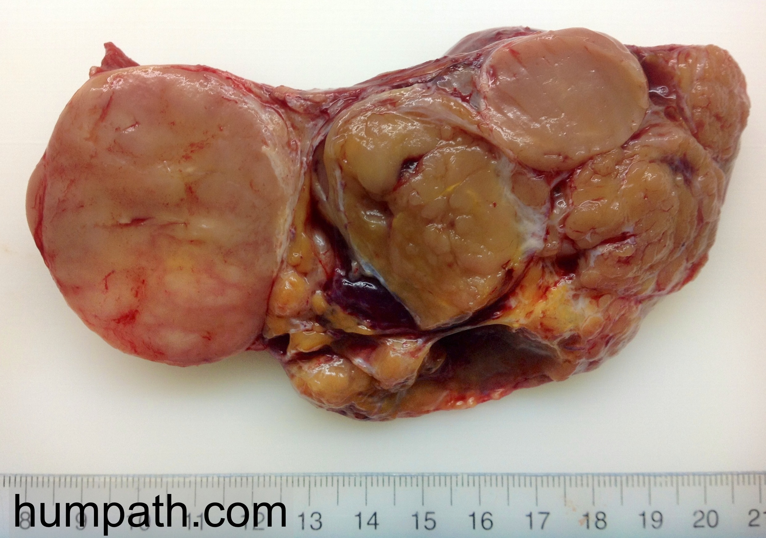
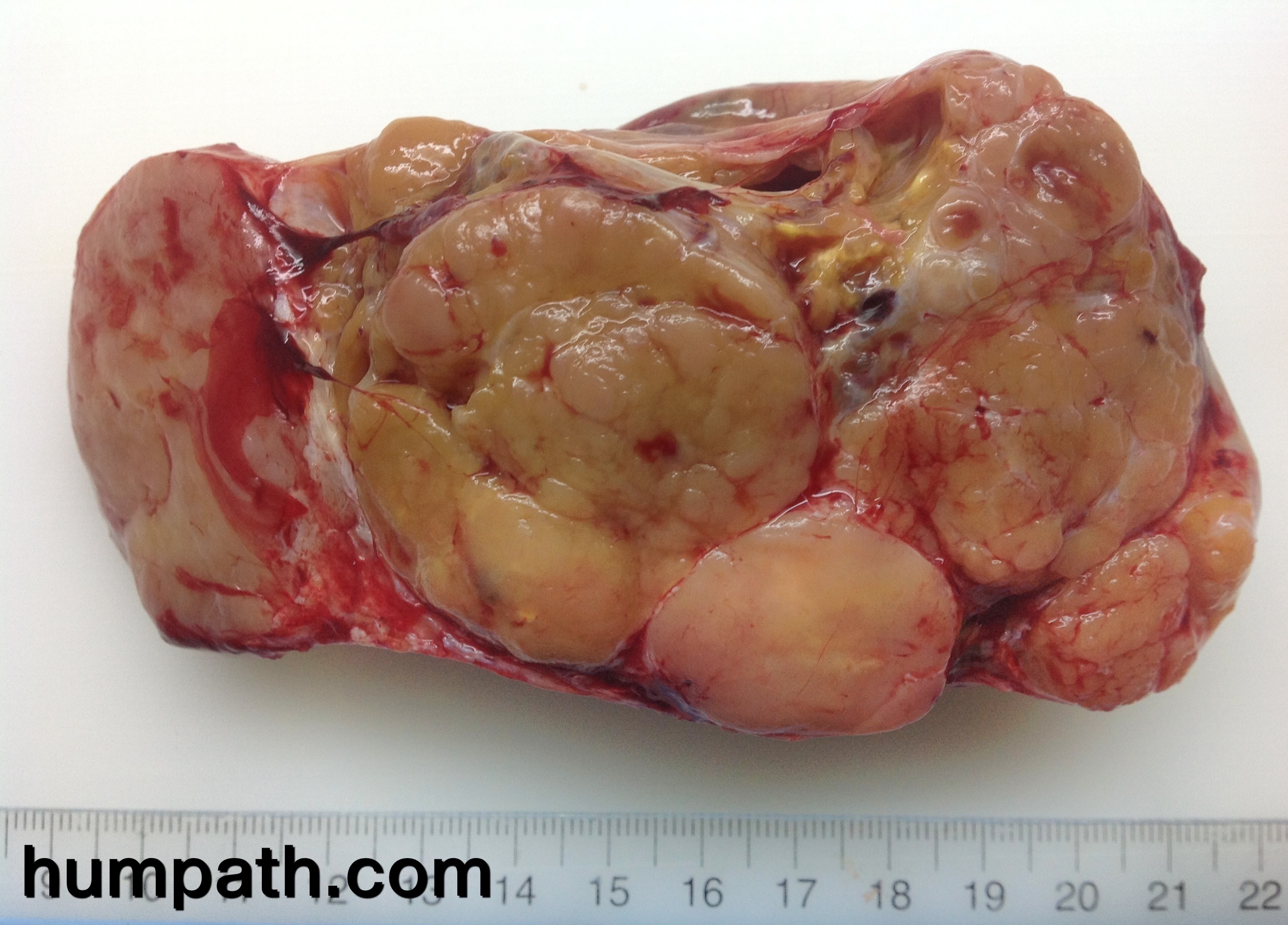
Type A thymomas generally have a thick fibrous capsule as shown here. Prominent fibrous bands course through the tumor separating it into lobules.
Type A thymoma is the least common subtype and accounts for 10%-12% of all thymomas.
The mean age at presentation is around 65 years which is slightly older than that for all thymomas.
Thymomas are large, multilobulated tumors. Note the fibrous septae dividing the tumor into variably-sized lobules.
About 17% of type A thymoma patients present with myasthenia gravis (muscle weakness, fatigue, diplopia, drooping eyelids, and dysphagia).
The remainder present with symptoms related to compression of anterior mediastinal structures (dyspnea, persistent cough, chest pain, fever, night sweats, and weight loss). Many are discovered incidentally on imaging studies performed for other reasons.
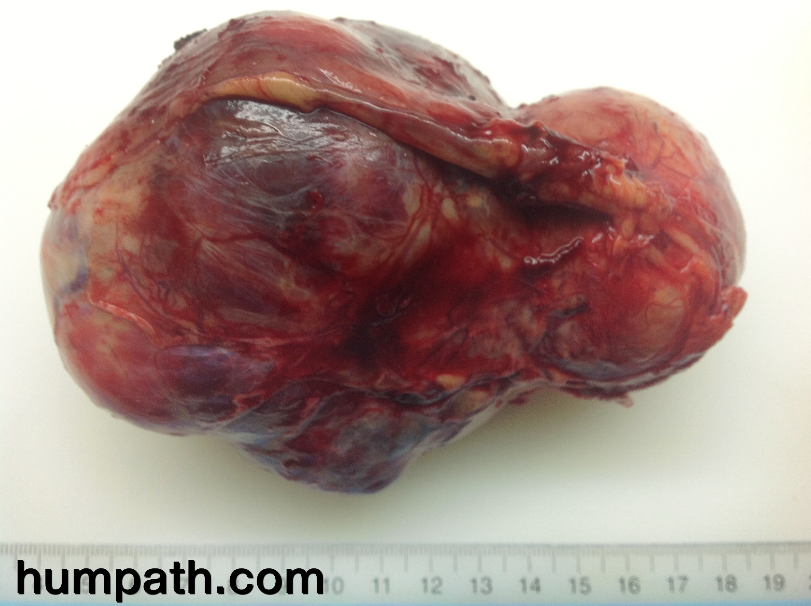
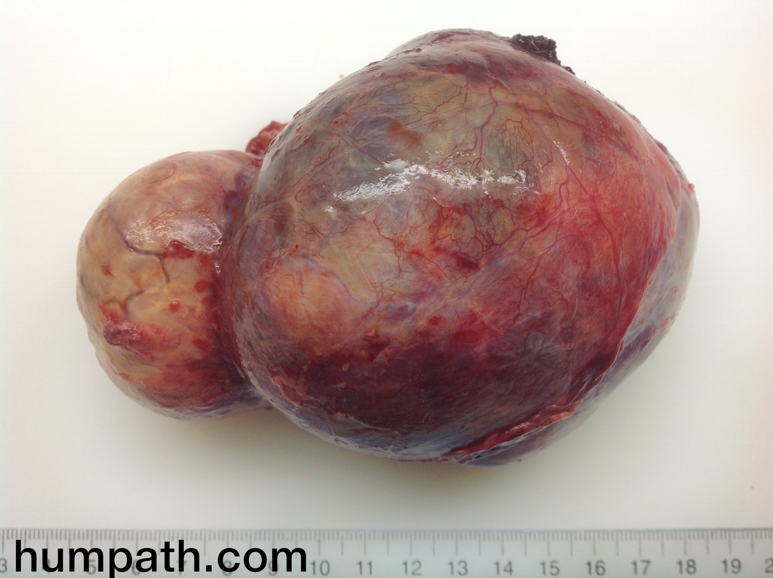
Most type A thymomas (about 60%) are diagnosed at Stage I (completely encapsulated) according to the Masaoka-Koga classification system.
About 30% of tumors are Stage II (microscopic capsular invasion OR gross invasion into surrounding fat OR adhesions to pleura or pericardium).
The image shows a bland spindle cell proliferation with a few scattered lymphocytes in the background.
Images
http://www.webpathology.com/image.asp?case=402&n;=1
Anterior mediastinal mass, encapsulated and non invasive - spindle cell thymoma (WHO type A)
Digital cases
JRC:3191 : Spindle cell thymoma. Thymoma, type A.
Microscopy
Type A thymoma is composed of bland spindle or oval epithelial cells admixed with few or no lymphocytes.
It can display a variety of architectural patterns, including microcystic foci, rosettes, glandular structures (shown here), meningioma-like whorls, storiform pattern, and hemangiopericytoma-like vascular areas. Multiple pattern are seen within the same tumor.
Type A thymoma is usually composed of spindle shaped cells arranged in short fascicles. They have oval or round bland nuclei with finely dispersed chromatin and inconspicuous nucleoli. Mitotic activity is low. A few gland-like structures with eosinophilic secretions can be seen on the left. There are only rare immature thymocytes scattered among the spindle cells.
If there are lymphoid aggregates OR greater than 10% of the tumor has moderate infiltrate of lymphocytes – such tumors are best classified as type AB thymomas.
Rare cases of type A thymoma show cytologic atypia, increased mitotic activity, and focal necrosis. The prognostic significance of this finding is not known.
See also
thymomas
- thymoma type AB
- thymoma type B