Home > E. Pathology by systems > Reproductive system > Male genital system > Prostate > Gleason pattern 3
Gleason pattern 3
Tuesday 31 July 2012
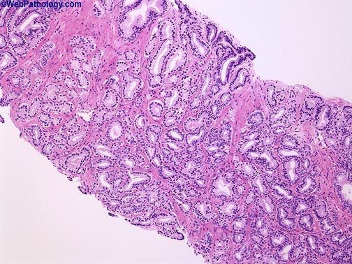
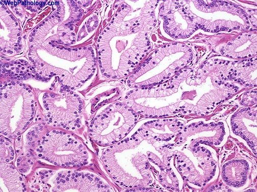
Gleason pattern 3 cancer consists of variably sized individual glands.
Individual cells
A departure from the original Gleason classification system is that “individual cells” would not be allowed within Gleason pattern 3.
Cribriform Gleason pattern 3
A further area of divergence from the original Gleason system is the controversial area of cribriform Gleason pattern 3.
Within Gleason’s original illustrations of his cribriform pattern 3, he depicts large cribriform glands, which the consensus panel would uniformly diagnose as cribriform pattern 4.
The consensus panel required extremely stringent criteria for the diagnosis of cribriform pattern 3, with remaining cribriform patterns typically falling into Gleason pattern 4.
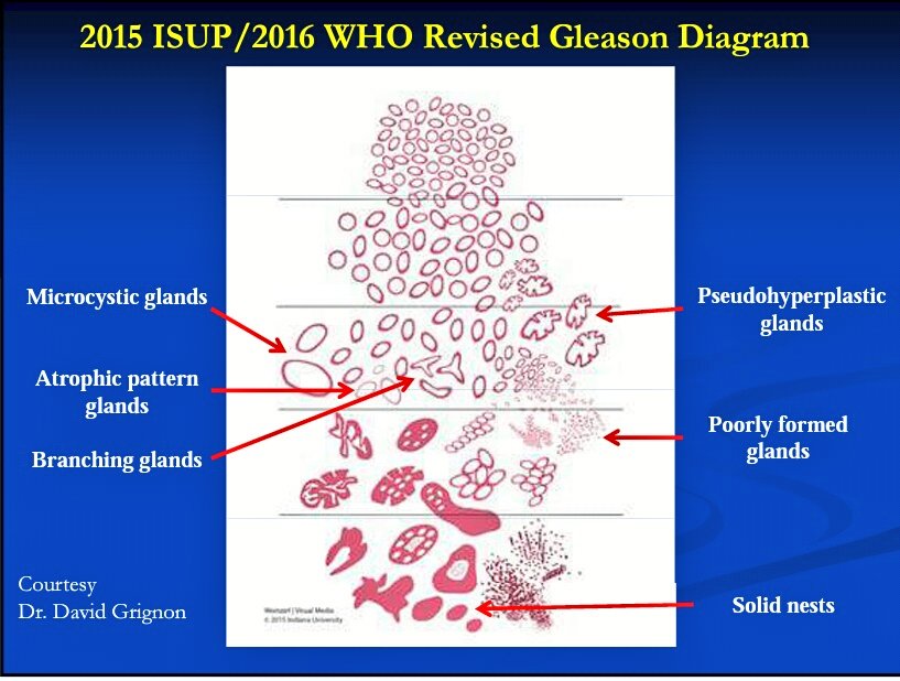
The criteria used to diagnose cribriform pattern 3 were rounded, well circumscribed glands of the same size of normal glands.
When various images were shown to the consensus panel of potential candidates for cribriform Gleason pattern 3, almost none of them met the criteria based on subtle features, such as slight irregularities of the outer border of the cribriform glands.
Subsequent to the 2005 ISUP meeting, JI Epstein reviewed 3590 consecutive prostate cancers sent to me over seven months; 30 needle biopsy cases were selected that possibly represented cribriform Gleason pattern 3 cancer, 36 digital images were taken and sent to ten experts in prostate pathology with a consensus defined when at least 7/10 experts agreed on the grade.
Even in this highly selected set of images thought to be the best candidates for cribriform pattern 3 from a busy consult service, most experts interpreted the cribriform patterns as pattern 4. There was only one consensus pattern 3 case.
Furthermore, most of the cribriform foci investigated (73%) were associated with more definitive pattern 4 elsewhere on the needle biopsy specimen.
There was poor reproducibility amongst experts as to cribriform pattern 3 vs. pattern 4 due to:
disagreement as to what are the key diagnostic features in a given case (i.e. irregular distribution of lumina and variable slit-like lumina, favor pattern 4 vs. small glands and regular contour, favor pattern 3;
disagreement as to assessment of given criteria: regular vs. irregular distribution of lumina and regular vs. irregular contour.
Conceptually, one would expect the change in grade from pattern 3 to pattern 4 to be reflected in a distinct architectural paradigm shift where cribriform as opposed to individual glands are formed, rather than merely a subjective continuum of differences in size, shape and contour of cribriform glands.
The only reason why cribriform pattern 3 even exists is because of the original Gleason schematic diagram.
Gleason never specifically published the prognostic difference between what he called cribriform Gleason pattern 3 compared to Gleason pattern 4.
Many of Gleason’s cribriform Gleason pattern 3 cancers may not even have been infiltrating carcinomas due to the lack of availability of immunohistochemistry for basal cell markers.
Today we might have diagnosed them either as cribriform high-grade PIN or intraductal carcinoma of the prostate (concepts not present in Gleason’s era).
Based on all the above data, all cribriform cancer should be interpreted as Gleason pattern 4 and not pattern 3.
See also
Gleason pattern 3
Gleason grading system