Home > E. Pathology by systems > Urinary system > allantois
allantois
Wednesday 25 November 2009
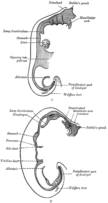
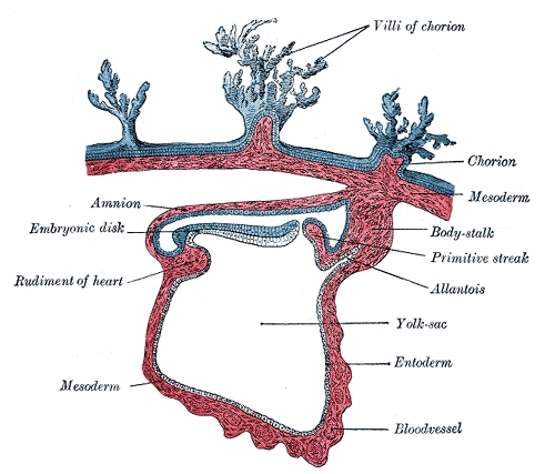
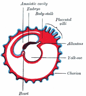
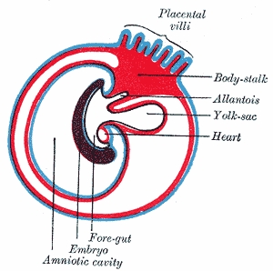
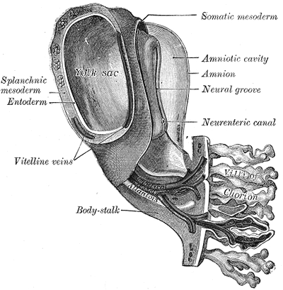
Allantois (plural allantoides or allantoises) is a part of a developing animal conceptus (which consists of all embryonic and extra-embryonic tissues). It helps the embryo exchange gases and handle liquid waste.
The allantois, along with the amnion and chorion (other embryonic membranes), identify humans as amniotes, along with reptiles, dinosaurs, birds, and other mammals. Of the vertebrates, only Ichthyopsidas (fish and amphibians) lack this structure.
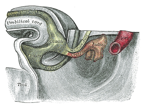
In placental mammals, the allantois is part of and forms an axis for the development of the umbilical cord.
The mouse allantois consists of mesodermal tissue, which undergoes vasculogenesis to form the mature umbilical artery and vein.
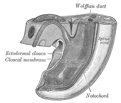
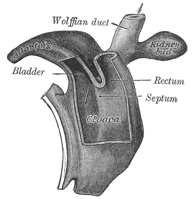
The human allantois is an endodermal evagination of the developing hindgut which becomes surrounded by the mesodermal connecting stalk. The connecting stalk forms the umbilical vasculature. These endodermal and mesodermal tissues together form the human umbilical cord. The allantois later definitively becomes the urachus, which removes nitrogenous waste from the fetal bladder.
Pathology
A patent allantois can result in urachal cyst.
Credits