Home > E. Pathology by systems > Locomotory system > Bones > metaphyseal fibrous defect
metaphyseal fibrous defect
Monday 9 March 2009
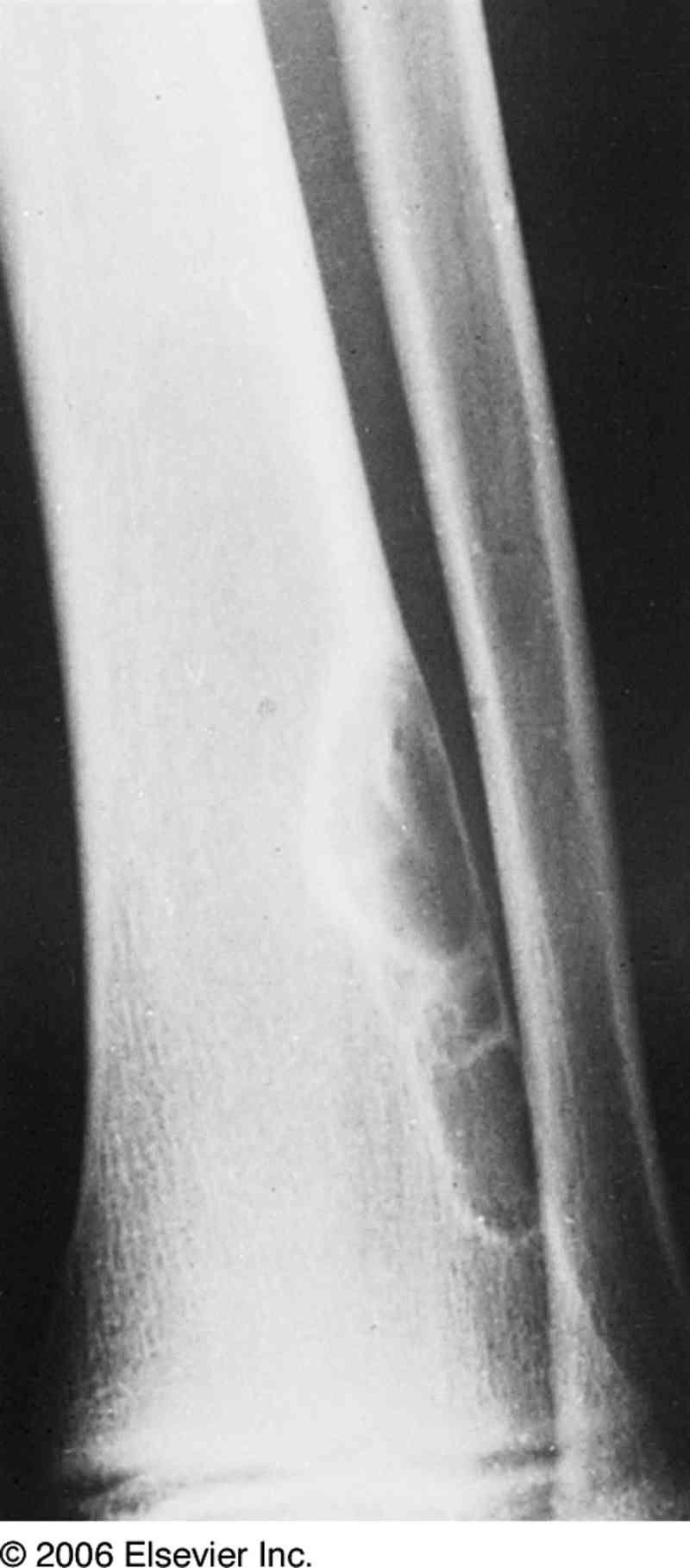
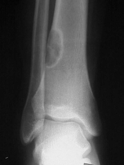
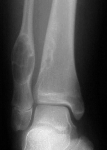
Definition: Eccentric, sharply delineated, metaphyseal lesion in long bones of adolescents. It can be considered as a variant of the lesional group associating fibrous cortical defect (FCD), non-ossifying fibroma (NOF) or and benign fibrous histiocytoma of bone (BFHB).
It can be associated with adamantinomas of long bones.
Nota bene: This lesion is called non-ossifying fibroma or benign fibrous histiocytoma if mass-forming and involving the medullary cavity.
Clinical Features
Adolescents
Few or no symptoms except pain
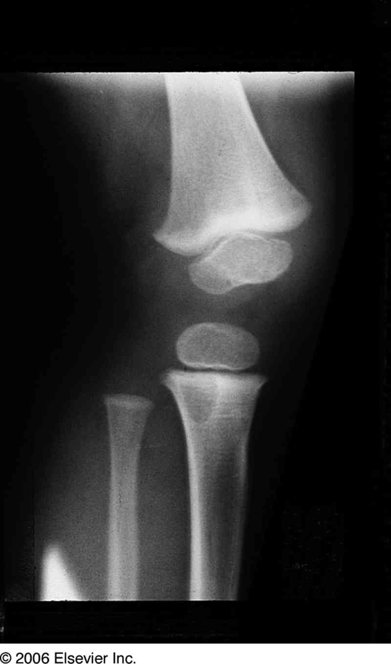
Usually found incidentally on radiography
Fractures can occur through the thinned cortex1
Pathogenesis
Controversy regarding whether neoplastic or developmental aberration at epiphyseal plate
Location:
usually long tubular bones, particularly: upper or lower tibia, lower femur
Macroscopy
Granular and brown or dark red
Eccentric
Sharply delimited
sharp delineation
sclerotic margins
Not too distant from epiphysis
Sometimes accompanied by epiphyseal disorders
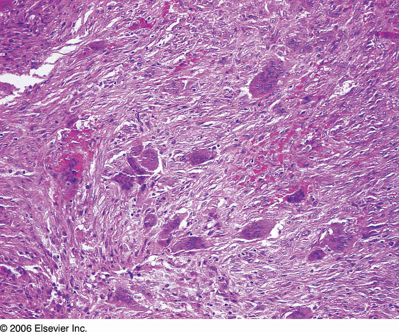
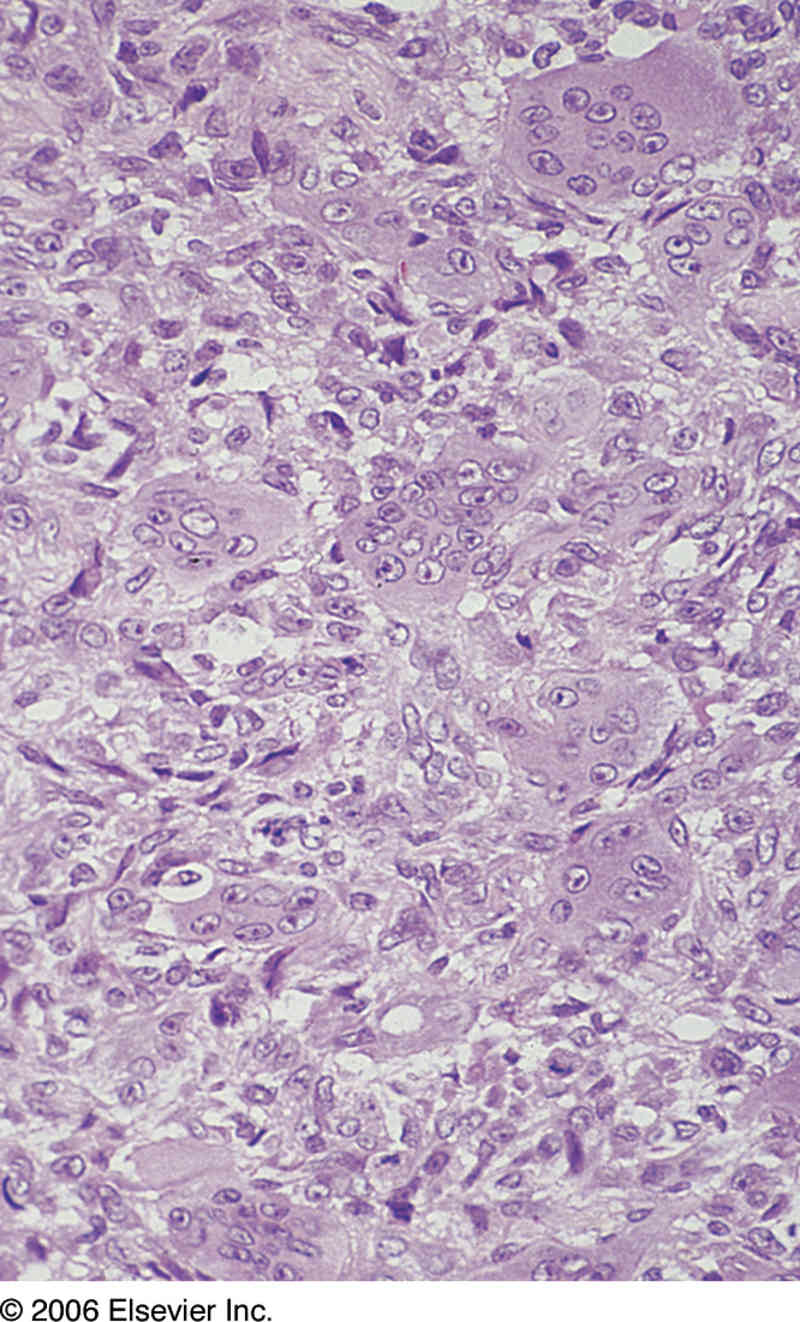
Histopathology
Cellular masses of fibrous tissue
often storiform pattern
spindle cell of fibroblastic appearance
irregularly scattered osteoclasts.
Frequent scattered osteoclasts and collections of foamy and hemosiderin-laden macrophages
Exceptionally, bizarre nuclear features, not necessarily indicative of malignant
Diagnosis
When loose and with an intramedullary component, designated non-ossifying fibroma or non-osteogenic fibroma
Differential Diagnosis
giant cell tumor of bone
malignant fibrous histiocytoma
aneurysmal bone cyst
chondroblastoma
Links
References