Home > E. Pathology by systems > Digestive system > Esophagus > esophageal duplication cyst
esophageal duplication cyst
Wednesday 15 October 2003
Digital cases
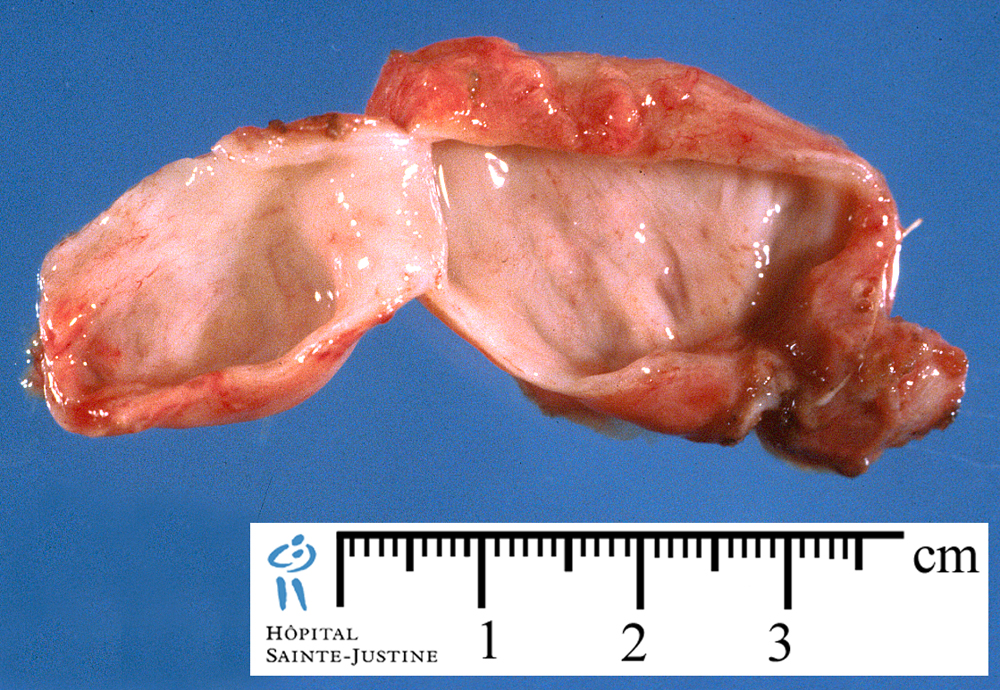
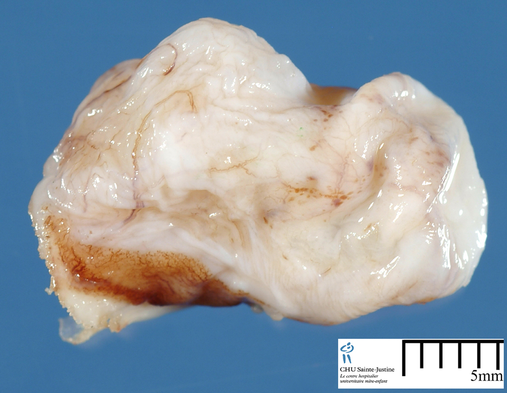
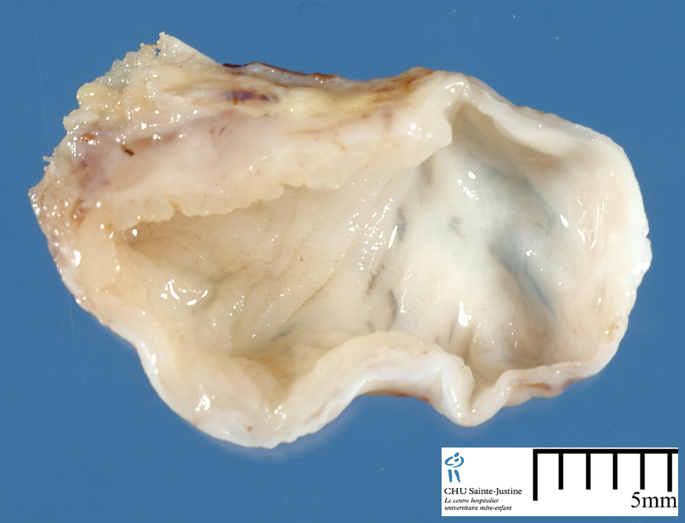
![]() Case 256 : Medial cervical digestive duplication cyst associated with mandibular anomalies
Case 256 : Medial cervical digestive duplication cyst associated with mandibular anomalies
Definition : The esophageal duplication cysts are thought to arise from foregut budding errors during the third to sixth week of gestation.
Clinical synopsis
The clinical diagnosis of an esophageal duplication cyst is made before the age of 2 years in 22% of the patients.
Duplication cysts in the mid and lower thirds of the esophagus are usually asymptomatic, and they are not usually accompanied by skeletal muscle layer.
Symptoms depend on the location of the esophageal duplication cyst and are generally caused by compression of adjacent structures.
Of those who are symptomatic, 70% present with dysphagia, 20% have epigastric discomfort, and 10% have retrosternal pain.
Respiratory distress is the most common clinical presentation of cervical
esophageal duplication cysts in infants.
Physiopathology
The esophageal duplication cysts are thought to arise from foregut budding errors during the third to sixth week of gestation.
During embryologic development, the laryngotracheal groove divides into the dorsal and ventral portions, which become the esophagus and respiratory tract, respectively.
An esophageal cyst forms when the secretory vacuoles during foregut luminal obliteration fail to coalesce. The timing of the budding error dictates the location of the cyst.
Early errors result in cysts being formed in the mediastinum such as cervical esophageal duplication cysts.
Localization
Esophageal duplication cysts can be tubular or spherical, and two thirds occur in the right side of the mediastinum caused by the dextrorotation of the stomach during embryogenesis.
Approximately 80% of cysts do not communicate with the esophageal lumen. The others generally run parallel to and communicate with the esophageal lumen.
In 60% of cases, the cyst is found in the distal portion of the esophagus.
Classification
Arbona et al. developed criteria for classifying a foregut duplication cyst as an esophageal cyst. These criteria include the following:![]() 1. The cyst is within or attached to the esophageal wall.
1. The cyst is within or attached to the esophageal wall.![]() 2. It is covered by 2 muscle layers.
2. It is covered by 2 muscle layers.![]() 3. The lining is squamous, columnar, cuboidal, psedostratified,
3. The lining is squamous, columnar, cuboidal, psedostratified,
or ciliated epithelium.
Furthermore, Palmer’s classification of esophageal cysts
requires the following:![]() 1. The cyst demonstrates attachment to the esophagus.
1. The cyst demonstrates attachment to the esophagus.![]() 2. Epithelium represents some level of the gastrointestinal
2. Epithelium represents some level of the gastrointestinal
tract.![]() 3. Two layers of muscularis propria are present.
3. Two layers of muscularis propria are present.
Antenatal diagnosis
Esophageal duplication cysts can also be diagnosed in the fetus on prenatal ultrasound examination. Kawahara et al. describe a case of an esophageal duplication cyst that was identified at 30 weeks gestation and managed surgically when the infant was 35 days of age because of stridor and feeding difficulties.
As with other cervical lesions (eg, teratoma, lymphangioma), an ex-utero intrapartum treatment (EXIT) procedure might be considered if high airway obstruction a duplication cyst is confirmed prenatally. However, no precedent for this has been found in the literature.
Malignancy
Malignancy within esophageal duplication cysts is rare but has been reported in the literature. For this reason, surgical resection is indicated for all symptomatic lesions and is favored for asymptomatic cysts to prevent future complications.
Surgery
Surgical options include cervical excision, axillary thoracotomy, median sternotomy, or a minimally invasive thorascopic approach based on the
location of the cyst in the mediastinum and the experience of the surgeon. Dissection of the cervical portion had to be accomplished
through the thoracic inlet.
Undoubtedly, a cervical resection would have been less technically challenging, with the only caveat being risk of injury to the recurrent laryngeal nerve.
See also
![]() primitive foregut cysts
primitive foregut cysts
- esophageal malformations
![]() digestive malformations
digestive malformations
- esophageal malformations
References
![]() Cervical esophageal duplication cyst: case report and review of the literature. Nayan S, Nguyen LH, Nguyen VH, Daniel SJ, Emil S. J Pediatr Surg. 2010 Sep;45(9):e1-5. PMID: 20850608
Cervical esophageal duplication cyst: case report and review of the literature. Nayan S, Nguyen LH, Nguyen VH, Daniel SJ, Emil S. J Pediatr Surg. 2010 Sep;45(9):e1-5. PMID: 20850608
![]() Wootton-Gorges S, Eckel G, Poulos ND, et al. Duplication of the cervical esophagus: a case report and review of literature. Pediatr Radiol 2002;32:533-5.
Wootton-Gorges S, Eckel G, Poulos ND, et al. Duplication of the cervical esophagus: a case report and review of literature. Pediatr Radiol 2002;32:533-5.
![]() Sodhi K, Saxena A, Rao K, et al. Esophageal duplication cyst an unusual cause of respiratory distress in infants. Pediatr Emerg Care 2005;21:854-6.
Sodhi K, Saxena A, Rao K, et al. Esophageal duplication cyst an unusual cause of respiratory distress in infants. Pediatr Emerg Care 2005;21:854-6.
![]() Moulton M, Moir C, Matasumoto J, et al. Esophageal duplication cyst: a rare cause of biphasic stridor and feeding difficulty. Int J Pediatr Otorhinolaryngol 2005;69:1129-33.
Moulton M, Moir C, Matasumoto J, et al. Esophageal duplication cyst: a rare cause of biphasic stridor and feeding difficulty. Int J Pediatr Otorhinolaryngol 2005;69:1129-33.
![]() Bishop HC, Koop CE. Surgical management of duplications of the alimentary tract. Am J Surg 1964;107:434-42.
Bishop HC, Koop CE. Surgical management of duplications of the alimentary tract. Am J Surg 1964;107:434-42.
![]() Arbona JL, Fazzi JG, Mayoral J. Congenital esophageal cysts: case report and review of literature. Am J Gastroenterol 1984;79(3):177-82.
Arbona JL, Fazzi JG, Mayoral J. Congenital esophageal cysts: case report and review of literature. Am J Gastroenterol 1984;79(3):177-82.
![]() Rhee R, Ray C, Kravetz M, et al. Cervical esophageal duplication cyst:MR imaging. J Comput Assist Tomogr 1988;12(4):693-5.
Rhee R, Ray C, Kravetz M, et al. Cervical esophageal duplication cyst:MR imaging. J Comput Assist Tomogr 1988;12(4):693-5.
![]() Sharma KK, Ranka P, Meratiya S. Isolated cervical esophageal duplication: a rarity. J Pediatr Surg 2005;40:591-2.
Sharma KK, Ranka P, Meratiya S. Isolated cervical esophageal duplication: a rarity. J Pediatr Surg 2005;40:591-2.
![]() Billmire DF, Allen JE. Duplication of the cervical esophagus in children. J Pediatr Surg 1995;30:1498-9.
Billmire DF, Allen JE. Duplication of the cervical esophagus in children. J Pediatr Surg 1995;30:1498-9.
![]() McCullagh M. Antenatal identification of a cervical oesophageal
McCullagh M. Antenatal identification of a cervical oesophageal
duplication. Pediatr Surg Int 2000;16(3):204.
![]() Gans SL, Poi-rs WJ. Anomalous lobe of lung arising from esophagus. Thoracic Surg 1951, 2!, 3 13-318.
Gans SL, Poi-rs WJ. Anomalous lobe of lung arising from esophagus. Thoracic Surg 1951, 2!, 3 13-318.
![]() Barzilai M, Sela L. Cervical esophageal duplication cyst a case report and review of the literature. Dig Surg 1995;12:199-202.
Barzilai M, Sela L. Cervical esophageal duplication cyst a case report and review of the literature. Dig Surg 1995;12:199-202.
![]() Winslow RE, Dykstra G, Scholten DJ, et al. Duplication of the cervical esophagus. An unrecognized cause of respiratory distress in infants. Am Surg 1984;50:506-8.
Winslow RE, Dykstra G, Scholten DJ, et al. Duplication of the cervical esophagus. An unrecognized cause of respiratory distress in infants. Am Surg 1984;50:506-8.
![]() Sherer D, Timor-Tritsch I, Dalloul M, et al. Prenatal sonographic findings of an isolated cervical esophageal duplication cyst. J Ultrasound Med 2009;28:405-7.
Sherer D, Timor-Tritsch I, Dalloul M, et al. Prenatal sonographic findings of an isolated cervical esophageal duplication cyst. J Ultrasound Med 2009;28:405-7.
![]() Kawahara H, Kamata S, Nose K, et al. Congenital mediastinal cystic abnormalities detected in utero: report of two cases. J Pediatr Gastroenterol Nutr 2001(33):202-5.
Kawahara H, Kamata S, Nose K, et al. Congenital mediastinal cystic abnormalities detected in utero: report of two cases. J Pediatr Gastroenterol Nutr 2001(33):202-5.
![]() Eichmann D, Engle S, Oldigs HD, et al. Radiological case of the month. Arch Pediatr Adolesc Med 2001;155:1067.
Eichmann D, Engle S, Oldigs HD, et al. Radiological case of the month. Arch Pediatr Adolesc Med 2001;155:1067.
![]() Snyder C, Bickler SW, Gittes GK, et al. Esophageal duplication cyst with esophageal web and tracheoesophageal fistula. J Pediatr Surg 1996;31(7):968-9.
Snyder C, Bickler SW, Gittes GK, et al. Esophageal duplication cyst with esophageal web and tracheoesophageal fistula. J Pediatr Surg 1996;31(7):968-9.
![]() Yasufuku M, Hatakeyama T, Maeda K, et al. Bronchopulmonary foregut malformation: a large bronchogenic cyst communicating with an esophageal duplication cyst. J Pediatr Surg 2003;38(2).
Yasufuku M, Hatakeyama T, Maeda K, et al. Bronchopulmonary foregut malformation: a large bronchogenic cyst communicating with an esophageal duplication cyst. J Pediatr Surg 2003;38(2).