Home > G. Tumoral pathology > cellular schwannoma
cellular schwannoma
Saturday 7 July 2007
Digital slides
JRC:357 : Cellular Schwannoma - Retroperitoneum.
JRC:96 : Cellular Schwannoma - Groin.
JRC:14942 : Cellular Schwannoma - Peritoneum (retroperitoenum).
JRC:485 : Cellular Schwannoma - Peritoneum.
JRC:15245 : Cellular Schwannoma - Peritoneum.
UPMC:518
Definition: The cellular variant of schwannoma (cellular schwannoma), mostly found in the deeper tissues, has been described.
Cellular schwannoma, described by Woodruff in 1981, is considered a variant of schwannoma and comprising about 10% of all schwannomas.
Localization
The most common location of cellular schwannoma is the paravertebral region with the sacral site constituting 64% of all neoplasms. The intracranial location accounts for 8% of all cellular schwannomas. The retrobulbar region is an extremely uncommon site for this tumor.
Microscopy
There are compact spindle-shaped cells with mitoses and some storiform areas, and a near absence of Verocay bodies and Antoni B tissue.
Rare variants with multiple glandular elements, with pseudoglandular structures, and with sweat duct differentiation have been reported.
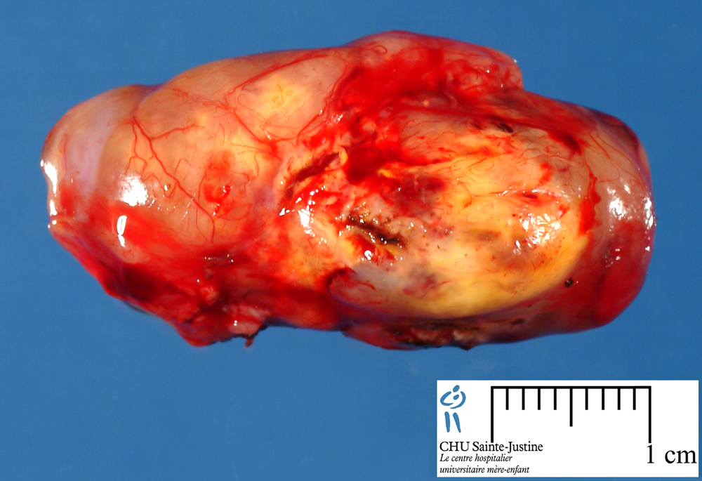
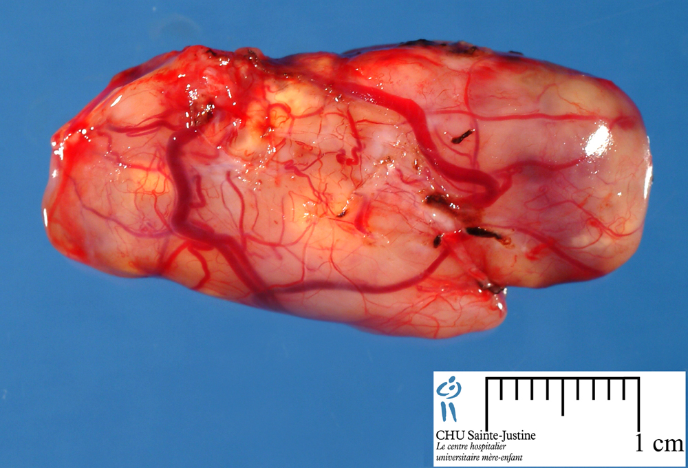
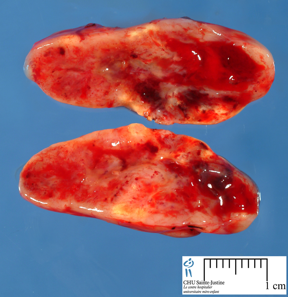
Cellular schwannoma is generally encapsulated and sometimes associated with a nerve. On histology, unlike classical schwannoma, cellular schwannoma discloses a markedly increase in cellularity, comprising fascicles of spindle cells which can occasionally be associated with herringbone or storiform pattern.
Compact and hypercellular fascicles recapitulating Antoni-A areas can be identified. Although classical Verocay bodies are seldom identified, there may be occasional suggestion of palisades. Antoni-B areas are, however, not prominently featured. The spindle cells may exhibit mild nuclear atypia. Thick-walled blood vessels usually displayed in classic schwannoma are also present in the cellular variant.
Microscopic areas of necrosis may be observed in cellular schwannoma, but geographic necrosis seen in malignant peripheral nerve sheath tumor (MPNST) is absent.
Mitotic activity usually does not exceed 4 per 10 high power fields although in plexiform cellular schwannoma in childhood, a mitotic index of up to 31 per 10 high power fields has been reported.
Immunochemistry
Cellular schwannoma is strongly positive for S-100 protein and vimentin. Glial fibrillary acidic protein (GFAP) is variably positive.
Ultrastructure
The striking feature seen ultrastructurally is the presence of numerous tightly packed intertwined cell processes as identified in our case. Primitive intercellular junctions and cell membranes abutting stroma and covered by well developed continuous and occasionally duplicated basement membranes, are also present. Luse bodies may be absent.
Differential diagnosis
malignant peripheral nerve sheath tumor (MPNST)
- due to its association with increased cellularity, nuclear atypia, mitotic activity, necrotic foci, bony erosion, and tumor recurrence.
- However, a MPNST is usually more cellular, associated usually with a higher degree of anaplasia, lacks the thick walled hyalinised blood vessels and usually demonstrates only focal S-100 positivity on immunohistochemistry.
In the orbital region, a solitary fibrous tumor has been confused for a schwannoma.
See also
schwannomas